
Oral Squamous Cell Carcinoma (OSCC) and Head and Neck Squamous Cell Carcinoma (HNSCC) have confounded the health care profession for a great length of time. A deadly and costly disease, the profession is still struggling with the ability to identify the disease in its earliest stages, when intervention is more likely to result in a favorable long-term prognosis.
According to the Oral Cancer Foundation, approximately 44,000 Americans will be diagnosed with oral or pharyngeal cancer this year, and 8,000 deaths will occur due to this disease. That is equivalent to one person dying every hour for an entire year. The five-year survival rate of those newly diagnosed with oral cancer has not significantly improved over the last 50 years.
Additionally, the cost ramifications of treatment after diagnosis can be quite staggering. It has been estimated that approximately $3.2 billion is spent in the United States alone every year in the treatment of these cancers.1
OSCC is an aggressive tumor with low response to chemotherapy and basic resistance to most standard of care anticancer drugs.2 The death rate of oral cancer is higher than that of many of the cancers that we routinely hear about: cervical cancer, Hodgkin’s lymphoma, testicular cancer and skin cancer. In fact, oral cancer is three times more common than cervical cancer with men having twice the likelihood of oral cancer as women. Additionally, oral cancer is the fourth leading cause of cancer in black men.
Who is at risk? Everyone! Twenty-five percent of oral cancer patients are non-smokers and non-drinkers.3 The other seventy-five percent of people have the classic risk factors including: smoking, using chewing tobacco, betel quid chewing and alcohol consumption. Individually these factors increase the risk of developing oral cancer, but when combined, the probability of disease development4 drastically increases. Studies show that there is a 16 to 36 percent chance of oral cancer reoccurrence, in addition to the probability of developing subsequent cancers elsewhere in the body.5,6,7
Although the oral cavity is easily accessible for examination and evaluation, there are several factors limiting the successful identification and early treatment of pre-malignant lesions. First, the current standard for screening and detection is a complex series of visual inspection and tactile palpation during an extra and intra oral examination by the health care professional; this is performed during routine dental or physical examination. The head and neck examination entails bimanual palpation of various external areas of: 1) the head and neck including the lower jaw, neck, glands and lymph nodes of the area, and 2) the oral cavity including the tongue, cheeks, floor and roof of the mouth, lips and back of the throat. Second, the ability to identify this disease in its earliest stages is not very easy and has often eluded the medical and dental professions. The reason? This disease can often be “occult” or hidden from plain view. Tissue that appears “normal” may often hide the truth within the cells below the surface of the mucosa.
If the disease is identified in Stage I or Stage II, which would be the ideal time for identification and before the dysplastic cells have been able to break through the basement membrane, the overall five-year survival rate is over 80%. All too often, however, the manifestations of this invasive and devastating disease are found in the late stage III or stage IV periods where the five-year survival rate falls to 20%. These “frank” lesions, when a lesion is apparent and easily and visually identified, are often are the hallmark in the identification process. Yet, when found at this stage, the lesion has typically advanced so deeply that it is impossible to treat without radical surgical intervention and significant loss of quality of life.
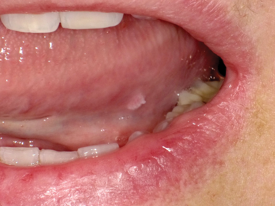
The need to identify these “hidden lesions” as early as possible and reduce the necessity for aggressive treatment and the ramifications that ensue, has promoted much research. Over the last 12 years many products have been developed to “visualize” or otherwise screen these early lesions, using a variety of techniques. While better than simple visual inspection with visible light and bimanual palpation alone, we must understand that these visualization modalities also present some challenges (Fig. 1).
FIGURE 1.
First, the operator must be able to clearly evaluate all structures in the oral and oropharyngeal cavity (Fig. 2). This task can often be difficult, especially with an uncooperative patient. Second, the resulting observation can often be misleading (Fig. 3). This is due to the subjective nature and interpretation of “results” obtained via the current visualization modalities with strong clinical research support lacking. As a result, lesion screening or visualization systems still do very little to address the clinical unmet need for early detection and intervention (Fig. 4).
FIGURE 2.

FIGURE 3.

FIGURE 4.

Only a “definitive test” can determine the biologic behavior (Fig. 5) of a lesion.9 Currently, that diagnostic “gold standard” for oral cancer diagnosis is histopathological examination of surgical biopsy specimens.10 Yet, if lesions are only biopsied when they are visible in the oral cavity (typically Stage III or Stage IV) we most certainly are not identifying disease at the earliest possible moment; this leads to late stage identification and lowering the long term prognosis and survivability of the patient.
FIGURE 5.

Clearly, a better mechanism is needed.
The National Institute of Dental and Craniofacial Research (NIDCR) created initiatives in 2002 for development of salivary diagnostic modalities for disease identification.11 Of the salivary biomarkers studied, only a few have been or are currently being translated for use in clinical practice.
Among the most promising biomarkers are protein measurements that include assessing total protein concentration and levels of CD44 (a cell surface transmembrane glycoprotein involved in cell proliferation and migration).12,13 CD44 is also a key tumor initiation marker14 that is over-expressed in the earliest stages of carcinogenesis.15,16 Soluble CD44 (solCD44) is released by proteinases and detectable in body fluids.17,18 It can be measured with simple, inexpensive assays.19-20

Research shows that the combination of solCD44 and total protein levels in oral rinses can distinguish OSCC cases from controls.20,21 This technology has been converted to a lateral flow test strip point-of-care and a laboratory test. The inclusion of a cancer stem cell marker will allow assessment of risk sufficiently early that reversal of carcinogenesis via behavioral change could be possible even before a lesion is clinically identified. This testing mechanism will be easy, quick, inexpensive and highly accurate. Most importantly, it will not interfere with the workflow of dental offices.
It is believed that the point-of-care test, with results easily obtained in minutes, will play a critical role in alerting and directing the clinician to action long before a lesion can be visibly detected. This testing mechanism, once available, will create a paradigm shift in our understanding of OSCC and how we classify, identify and treat it in the future.OH
John C. Comisi is a Private General Practice Dentist in Ithaca, NY and serves as the Dental Liaison consultant for Vigilant Biosciences Inc., a leading innovator and developer of solutions that aid in the early detection and intervention of cancer.
Oral Health welcomes this original article.
COMISI REFERENCES:
1. http://www.oralcancerfoundation.org/facts/
2. Martin Grimm, Marcel Cetindis, Max Lehmann, Thorsten Biegner, Adelheid Munz, Peter Teriete, Wiebke Kraut and Siegmar Reinert. Association of cancer metabolism-related proteins with oral carcinogenesis – indications for chemoprevention and metabolic sensitizing of oral squamous cell carcinoma? Journal of Translational Medicine 2014, 12:208 doi:10.1186/1479-5876-12-208
3. Llewellyn CD, Linklater K, Bell J, Johnson NW, Warnakulasuriya KA (2003). Squamous cell carcinoma of the oral cavity in patients aged 45 years and under: a descriptive analysis of 116 cases diagnosed in the South East of England from 1990 to 1997. Oral Oncol 2003; 39:106–114.
4. Ragin CCR, Modugno F, Gollin SM. The Epidemiology and Risk Factors of Head and Neck Cancer: a Focus on Human Papillomavirus. J DENT RES February 2007 86: 104-114
5. Schwartz LH, Ozsahin M, Zhang GN, et al. Synchronous and metachronous head and neck carcinomas. Cancer 1994;74:1933-8.
6. http://www.oralcancerfoundation.org/facts/metastasis.php
7. Hong WK, Lippman SM, Itri LM, et al. Prevention of secondary primary tumors with isotretinoin in squamous cell carcinoma of the head and neck. N Engl J Med 1990;323:795-801, 8257.
8. http://www.oralcancerfoundation.org/discovery-diagnosis/stages-of-cancer.php
9. R. Mehrotra, M. Hullmann, R. Smeets, T. E. Reichert, and O. Driemel, “Oral cytology revisited,” Journal of Oral Pathology and Medicine, vol. 38, no. 2, pp. 161–166, 2009.
10. Retham MP, Carpenter W, et al. Evidence Based Clinical Recommendations Regarding Screening for Oral Squamous Cell Carcinomas. J Am Dent Assoc 2010; 141: 509-520.
11. Wong DT. Salivary diagnostics powered by nanotechnologies, proteomics and genomics. J Am Dent Assoc 2006; 137: 313-321.
12. Ponta H, Sherman L, Herrlich PA. CD44: from adhesion molecules to signaling regulators. Nature Rev Mol Cell Biol. 2003;4:33-45.
13. Perez A, Neskey DM, Wen J, et al. CD44 interacts with EGFR and promotes head and neck squamous cell carcinoma initiation and progression. Oral Oncol. 2013;59(4):306-313.
14. Prince ME, Sivanandan R, Kacsorowski A, et al. Identification of a subpopulation of cells with cancer stem cell properties in head and neck squamous cell carcinoma.Proc Natl Acad Sci USA. 2007;104:973-978
15. Hirvikoski P, Tammi R, Kumpulainen E, et al. Irregular expression of hyaluronan and its CD44 receptor is associated with metastatic phenotype in laryngeal squamous cell carcinoma. Virchows Arch. 1999;434(1):37-44.
16. Ioachim E, Assimakopoulos D, Goussia AC, et al. Glycoprotein CD44 expression in benign, premalignant and malignant epithelial lesions of the larynx: an immunohistochemical study including correlation with Rb, p53, Ki-67 and PCNA.Histol Histopathol. 1999;14:1113-1118.
17. Dasari S, Rajendra W, Valluru L. Evaluation of soluble CD44 protein marker to distinguish the premalignant and malignant carcinoma cases in cervical cancer patients. Med Oncol. 2014 2014;31(9):139.
18. Kajita M, Itoh Y, Chiba T, et al. Membrane-type 1 matrix metalloproteinase cleaves CD44 and promotes cell migration. J. Cell Biol. 2001;153(5):893-904.
19.Franzmann EJ, Reategui EP, Carraway KL, et al. Salivary soluble CD44: a potential molecular marker for head and neck cancer. Cancer Epidemiol Biomarkers PrevMarch 2005;14;735.
20. Franzmann EJ, Reategui EP, Pedroso F, et al. Soluble CD44 is a potential marker for the early detection of head and neck cancer. Cancer Epidemiol Biomarkers Prev2007;16(7):1348-1355.


