
Abstract
Digital dentistry has existed for over 30 years, but many dentists are still reluctant to incorporate this technology into their practice. To many colleagues, digital techniques are only a marketing tool to impress patients. Yet, digital technology provides us with ways and means to improve and facilitate our dental procedures. Dental laboratories, for their part, have long incorporated these technologies; this digital shift allows them to be more efficient and to improve the quality of their work.
Introduction
Today, in most dental practices, patient’s interest in cosmetic procedures and smile enhancement is a reality. Once basic restoration and periodontics care are completed, patients often ask us how possible it would be for them to have a smile like those of the artists they see on television and in the movies.
Most patients want to have white and straight teeth, and wrongly think that getting the smile of their dream is a relatively easy task. Our responsibility as a dentist is to explain them in detail the different treatment options. This multidisciplinary approach with the help of our specialist colleagues (orthodontists, periodontists, maxillofacial surgeons, dental technicians) is often necessary in order to obtain the optimal result. Apart from purely dental health considerations, aesthetic treatment has an important psychological factor. Each patient has personal reasons to improve their smile. Our role is to take the needed time to listen carefully to their rationale and expectations. Starting from there, one must suggest a treatment plan with different options, and obtain the informed consent of the patient before starting any procedure.
In the Internet age and social networks, patients have access to a lot of information, and often they come to our office with a list of claims, and have a more or less well-informed idea of the treatment they require. Our role as a health professional is to explain in detail the advantages and inconvenient of each treatments so that the patient makes an informed choice according to his condition.
There are many ways today to demonstrate to a patient the possibilities of aesthetic dentistry. Several programs from different companies allow us from a photo, to change the shape and position of the teeth, and show a photo “before/after” treatment. On the other hand, these tools have their limitations and can create unrealistic expectations. More often than not, it is impossible to recreate that artificially created smile in the end. We must not transfer our obligation of means into an obligation of result. Nothing is more frustrating than finishing a case of aesthetics and having a patient disappointed with the results. When this happens, a communication and planning problem at the beginning is the source.
A dentist should, in any elective treatment involving aesthetic, require a complete examination with radiographs, casts, and photographs. These pre-op photographs are used for planning, and also as a basic reference to demonstrate to the patient their initial situation. Patients often forget their initial condition, and these pictures can help the dentist in cases where the patient is not satisfied with the final result.
The production of a diagnostic wax model is a must for each of these cases. This is a task that should be done by the dentist himself or a dental technician to whom the patient’s expectations are communicated. Waxing is a long and complex work when carried out by the conventional technique. Often, for these reasons, dentists do not care to do it; they thus let the technician decide the final outcome, without having the proper information to give the optimum result. This attitude can lead to a disappointing end result or expensive laboratory alterations.
CASE #1: Female Patient, age 59
This patient consults, because she is dissatisfied with her smile. Her medical condition is good; she postponed dental treatments due to anxiety and poor dental experiences.
Fig. 1
Pre-op photo taken at initial consultation.

Fig. 2
Patient’s Initial Smile.

Fig. 3
Using the Initial Photo at Waxing Stage.

Fig. 4
Superimposing Original Photo and Diagnosis Wax model.

Fig. 5
Upper and Lower Maxillaries Diagnostic Waxing From Digital Impression and Initial Photography.

Fig. 6
After Treatment Smile.

Fig. 7
Upper Teeth Restored (1.3 to 2.3).

CASE #2: Patient age 60
Medicated with Bisphosphonate for osteoporosis onset. Noticeable wear of anterior teeth and a number of defective restorations. Subpar aesthetics. Canine and anterior guide inexistent.
Fig. 1
Pre-op photo taken at initial consultation.

Fig. 2
Using Superimposition of Photos for Waxing. Notice the red dots for interpupillary line and the blue dot for median line.

Fig. 3
Machined PMMA (polymethylmethacrylate) Matrix from Diagnostic Wax Model.
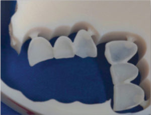
Fig. 4
Digital Waxing.

Fig. 5
Completed Lab Finished Matrixes.

Fig. 6
Trial of a PMMA Matrix.

Fig. 7
Trial of Both PMMA Matrix.

Fig. 8
Final Permanent Restoration.

Fig. 9

3D Planning Digital Technique
The technique we have developed uses digital technology in the office and in the laboratory. When the patient consults us for cosmetic dentistry, our examination includes digital radiographs, high-quality digital pictures with specialized equipment, and digital impressions of the two mouth arcades with digital articulation.
The photo of the patient’s face should be taken upright, and the patient should make his smile complete in order to allow a correlation with the digital photo (.JPG) and the file of the digital impression (.STL). We also take close-up pictures with the help of mirror and retractors to complete our photo documentation.
From the (.STL) file obtained with the digital impression (3M True Definition Scanner) and JPEG-quality photos, the laboratory can quickly produce a diagnostic wax model using its own drawing software such as (3 Shape Design Center). All these systems have open platforms, allowing their compatibility. The diagnostic wax model should take into account the occlusion, as well as the patient’s facial parameters such as the interpupillary line, median line, smile line and position of the lip. When finished, the technician will send us a proposal that can easily be altered, should it become necessary.
The next appointment with the patient is to explain our detailed treatment plan. Using documentation at different stages, the explanation to the patient becomes very clear. They can then make an informed decision about their treatment and accept or refuse to proceed accordingly.
Should the patient accept the treatment plan, a machined CAD/CAM-model, and a polysiloxane matrix will be used to prepare the temporary restoration. In certain situations, when the occlusion permits, it is also possible to make a matrix machined in PMMA (polymethylmethacrylate), which can be tried in the mouth of the patient. This is the best way for the patient to visualize the change anticipated by our future treatments.
The marked advantage of this digital technique is that we can plan our processing in a very precise way, and simplify the realization. The cost of this planning is very reasonable because the dental technician quickly produces the digital model. This technology reduces costs for the dentist and for the patient as well.
Finally, the advantage of this temporary is that it allows us to check all our parameters directly in the mouth before the realization of the permanent restorations. If the temporary is adequate, it can be scanned again to provide relevant information to the technician. Otherwise, you can change the temporary restoration by chair side, and then re-scan. All these steps are quickly done digitally, and facilitate control all the phases of most complex treatments.
Conclusion
The integration of digital technology is an undeniable reality in dentistry. If major dental laboratories have achieved this integration, dentists must acknowledge all the benefits that these changes can bring to their day-to-day practice. Modern digital technologies, using multiple software and open platforms, simplify our work and improve treatments provided to our patients. OH
Oral Health welcomes this original article.
Acknowledgement
Mr. Marc Nantais, owner of LAB LONGUEUIL (info@lablongueuil.com) for dental laboratory support. Paul Trépanier, DMD, for review and translation.
About the Author
Dr. Marc Robert graduated from the University of Montréal in 1988. As a general practitioner practicing in the Montreal area (Longueuil), he has successfully integrated several technologies (laser and CAD / CAM) over the years. He teaches senior year students at the University of Montréal a course on laser introduction and CAD / CAM (Planmeca Fit and 3M True Definition Scanner). He is also an instructor in laser (Biolase) and CAD/CAM (Planmeca Fit) for his colleague’s dentists. Members of multiple national and international dental organizations, he is a Fellow of the American College of Dentists. His direct speech, candor, and simplicity make him a popular speaker. Dr. Robert was the dentist of the first two seasons of the TV shows What Age Do You Give? (Ten Years Younger) produced by the Canal Vie television network in Quebec.
RELATED ARTICLE: Accuracy and Resolution in 3D Digital Scanning
Follow the Oral Health Group on Facebook, Instagram, Twitter and LinkedIn for the latest updates on news, clinical articles, practice management and more!


