Pediatric dentistry has seen considerable improvements in materials and treatment options over the past few years. While other dental specialties have experienced tremendous developments in their materials and methods over the past forty years, pediatric dentistry has seemed to evolve much more slowly. Although we have enjoyed success with the treatments of the past, less invasive options involving fewer and more biocompatible chemicals can only benefit our patients and our practice of dentistry. What is good for our patients is good for our profession. This article will focus on the following new developments in pediatric dentistry: less invasive pulpal treatment, the switch from formocresol to MTA, bioactive restorations, and silver diamine fluoride.
I was trained in pediatric dentistry from 2005-2007. At the time, I had a very experienced clinical professor who felt that we needed to perform complete caries excavation and a therapeutic pulpotomy if we were very near the pulp. That thought process had been around for many years – it was just the way it was always done. Now that numerous studies have emerged stating that indirect pulp therapy is more successful than therapeutic pulpotomies, 1 practitioners are comfortable with performing this less invasive procedure in the absence of signs or symptoms of irreversible pulpitis.
When a pulpotomy is indicated, the majority of practitioners that I speak with are using formocresol, a rather controversial medicament. Formocresol has been used for decades in pulp therapy with many studies indicating that it is safe if used the correct way. 2 However, it is a “toxic chemical” with a “bad rap” when looked at through the public’s eye. My thought process is simply this: if we can use a material that provides equal or better outcomes 3 and is less toxic, why shouldn’t we? There are two primary alternatives to formocresol for therapeutic pulpotomies: ferric sulfate and Mineral Trioxide Aggregate (MTA). There are ample studies in the literature that show ferric sulfate has an equal success when compared to formocresol; however, it has a reputation among many users that it leads to a higher incidence of internal resorption. A lot of practitioners made the switch to ferric sulfate, became frustrated at seeing radiographic failure, and are going back to their tried and true formocresol.
When considering MTA, the number one reason that practitioners are not using it, is COST. Now that there are cost effective MTA options, we should all consider switching. Research shows that MTA has the highest success rate of any material used in pulp therapy. Perhaps the next most important benefit of using MTA in pulpotomies is the fact that it forms the ultimate protective pulpal seal and can be followed with more cosmetic options when compared to stainless steel crowns. Simply put, the enemy of primary tooth pulp therapy is leakage; therefore, stainless steel crowns have long been recommended following pulp therapy. Study after study shows that formocresol pulpotomies have a much higher success rate when followed by stainless steel crowns vs. other restorative options. With the use of MTA, the practitioner can feel more comfortable restoring select teeth with their material of choice. Biodentine by Septodont is an MTA substitute which can be used in therapeutic pulpotomies. NeoMTA by NuSmile is an MTA that is the least expensive MTA on the market. There are many great references available if the practitioners do their due diligence before choosing a material. I urge readers to look at MTA as another option.
Case 1
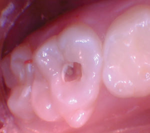
An 8-year-old patient presented with DO decay in a first primary molar exhibiting symptoms of reversible pulpitis. The tooth was treatment planned for a DO restoration with Activa Bioactive Restorative (Pulpdent). Upon decay excavation, a pulp exposure occurred necessitating a therapeutic pulpotomy. Due to the patient’s age, I felt comfortable proceeding with a therapeutic pulpotomy with Biodentine (Septodont) followed by an esthetic restoration. If the patient had been four to five years of age, I would have restored with a stainless steel crown due to its proven track record of longevity.
Improvements in resin-based composite materials now provide practitioners with “active” materials and bulk fill options that offer increased efficiency and predictability when restoring decayed teeth. It is important to remember that traditional composites are inert, passive materials. The modern day diet – high in acidic drinks and other sugar-laden snacks–presents a challenge that swings the pendulum in favor of recurrent decay. When a practitioner considers restorative options for primary teeth, dietary habits and oral hygiene must be considered.
Glass ionomers and resin modified glass ionomers have long been alternatives to resin-based composites due to their high fluoride release for teeth susceptible to recurrent decay. While GIs and RMGIs have proven benefits, they also lack certain characteristics that are desirable in pediatric restorations – longevity, compressive strength, and fracture resistance. Pulpdent has introduced an alternative with their Activa Bioactive product range. Activa is a urethane-based material that contains no Bisphenol A and no BPA derivatives. It releases and recharges calcium, phosphate and fluoride, which are invaluable for remineralization and preventing recurrent decay. A unique physical property of Activa is the very high fracture resistance resulting from a patented rubberized-urethane molecule inserted into the resin matrix. The compressive strength is comparable to traditional resin-based composites. So think of Activa as a material with bioactive properties greater than GIs and RMGIs, and physical properties comparable to traditional resin-based composites.
Fig. 1
Pre-operative view of upper first primary molar.

Fig. 2
Pulpal exposure occurred when excavating infected dentin (pulp exposure not captured in the photo).

Fig. 3
After unroofing the pulpal chamber.

Fig. 4
After the pulpal tissue was excavated to the canal orifices, a dry cotton pledget was used to assess pulp health by applying pressure hemostasis.

Fig. 5
This pulp exhibited a healthy radicular pulp as evidenced by easily controlled bleeding.

Fig. 6
2-3 mm layer of Biodentine was placed followed by a layer of Ionoseal (Voco) to prevent washout of MTA when proceeding with the restorative process.

Fig. 7
After acid etching and using bonding agent Scotchbond Universal (3M), the two upper primary molars were restored with Activa Bioactive Restorative A2.

Case 2
A 7-year-old new patient presented with significant decay and hypocalcification in the upper right first permanent molar. Decayed and hypocalcified first permanent molars in pediatric patients have long been proven to give the practitioner a difficult restorative dilemma. Should we attempt to prepare and restore conservatively? Should we be more aggressive and restore with full coverage such as a well-adapted stainless steel crown? Activa provides a great alternative in these cases, with calcium, phosphate and fluoride release coupled with high strength and fracture resistance.
Fig. 1
After acid etching and using bonding agent Scotchbond Universal (3M), the two upper primary molars were restored with Activa Bioactive Restorative A2.

Fig. 2
Once the decay was excavated and margins placed on solid surfaces, a heavy bevel was placed in the enamel to increase bond strength and marginal integrity. The central dark area was solid. The tooth was lined with Activa base/liner.

Fig. 3
After selective etching and using Scotchbond Universal, the tooth was restored with Activa Restorative A2.

Fig. 4
18-month follow up showed no fractures and intact margins.

Case 3
Recurrent decay in a primary tooth involving multiple surfaces usually leads straight to full coverage and rightfully so. We know that resin based materials leak in harsh acidic and sucrose riddled environments; therefore, full coverage is highly recommended in these cases. What if we could restore with a more esthetic material for the esthetic driven parents? In these esthetic cases, we still need strength and a non-inert, active material.
Fig. 1
Pre-operative photo of upper right 2nd primary molar with disto-occluso-buccal decay. In the past, for me at least, the treatment would have been a full coverage crown (likely stainless steel). Due to esthetic concerns from parents, it is nice to have an alternative option, especially a material that is active within the oral environment.

Fig. 2
The decay was excavated and all undermined enamel was removed.

Fig. 3
A band and wedge were placed. Activa Bioactive Restorative A2 was placed, cured, finished and polished.

Case 4
This 16-year-old male had a post-orthodontic relapse of a midline diastema and was self-conscious about the space. Options for closing the space were given, including six months of orthodontic correction vs. restorative correction with direct restorations. Due to the long, slender shape of his central incisors, the patient, his mother and I agreed that the best option would be restorative correction.
The introduction of Advantage Arrest with silver diamine fluoride (SDF) by Elevate Oral Care has provided a medicinal approach to treating carious lesions. SDF is a silver based fluoride liquid used to arrest carious lesions by denaturing and breaking down bacteria in the infected lesion. What makes SDF an adjunct to restorative care is its ability to penetrate dentinal tubules and prevent sensitivity in deep lesions when used in indirect pulp therapy. SDF, when used alone, has the ability to arrest incipient lesions. Arresting these lesions has the potential to eliminate the need for treatment under general anesthesia or restorative care in young children who are not able to cooperative in a normal dental setting. In my experience, SDF has definite limitations and is not a “silver bullet”. Depth of penetration is an issue in deep lesions with little access to the full extent of the lesion – those cases with a small access opening that expand and “balloon out” at the DEJ. If the lesion is into dentin, the depth of the lesion needs to be fully exposed, for SDF to truly arrest the decay.
Fig. 1
Pre-operative photo of a 2.75 mm diastema in a concerned teenager (post orthodontic relapse).

Fig. 2
After placement of two Bioclear matrices, the teeth were lightly beveled, acid etched and bonded with Scotchbond Universal. Activa Bioactive Restorative A2 was placed and cured. The teeth restorations were then finished and polished.

Fig. 3
When using Activa, I’ve noticed a blending, chameleon nature of the material. It typically blends beautifully with enamel when placed as a one shade restoration in the anterior. A fixed 1-1 retainer was placed after the restorations to prevent additional relapse.

Case 5
A 13-year-old female presented for a new patient exam. A large carious lesion was noted on upper left first permanent molar, which exhibited symptoms of reversible pulpitis. Treatment options were discussed with the patient and her mother. My personal goals for this case were to avoid a pulp exposure and to restore form and function.
Fig. 1
Initial clinical presentation of upper left 1st permanent molar with occlusal decay.

Fig. 2
When the lesion was unroofed, it was evident that a broad, deep lesion was present.

Fig. 3
With optimal convenience form achieved, the peripheral walls and DEJ were thoroughly excavated. Affected dentin was remaining on the pulpal floor to avoid a pulp exposure.

Fig. 4
A microbrush was used to apply silver diamine fluoride to the affected dentin.

Fig. 5
Ionoseal was placed on the pulpal floor and cured which accelerated the exposure of the silver ion precipitate (as evidenced by the black deposits).

Fig. 6
Activa Bioactive Restorative was placed, cured and finished. The SDF discoloration showing through the restoration, is definitely an esthetic compromise; however, the benefits of SDF in this case should provide longevity and restore form and function for years to come. In hindsight, an opaquer could have been used to mask the silver stain making the restoration more esthetic.

Case 6
A 2-year-old presented with carious lesions on all of her primary molars. Treatment options were discussed with the patient’s primary caregiver which included: improved home-care only, general anesthesia for restorative care or treatment with silver diamine fluoride. The patient’s caregiver chose silver diamine fluoride.
Fig. 1
Post SDF presentation of the lower right primary molars showing arrested lesions.

The oral environment is harsh, especially with the high acidic and high sucrose diets that we are seeing today in our offices. As materials continue to advance, it is crucial to our patients that we continue to evolve and provide the most advanced minimally invasive materials available. As dental materials improve, so should our oral health education to our patients. With the addition of these materials, we have restorative treatment options that we never had before. This is a win-win for us and for our patients. OH
Oral Health welcomes this original article.
References
1. Farooq, NS, et al. Success rates of formocresol pulpotomy and indirect pulp therapy in the treatment of deep dentinal caries in primary teeth. Pediatric Dentistry 22:4: 278-86, 2000.
2. Milnes, A. Persuasive Evidence that Formocresol Use in Pediatric Dentistry is Safe. J Can Dent Assoc 72(3): 247-8, 2006.
3. Peng, L, et al. Evaluation of the formocresol versus mineral trioxide aggregate primary molar pulpotomy: a meta-analysis. Oral Surg Oral Med Oral Pathol Oral Radiol Endod. 2006 Dec;102(6): e40-4.
About the Author
Dr. Wren is the founder of Wren Pediatric Dentistry and is now co-owner of Wren and Bryant Pediatric Dentistry in Brandon, Mississippi. Dr. Wren is the founder of Pediatric Dental Seminars which serves to educate general dentists on topics related to pediatric dentistry through lecture-based seminars and hands on workshops.
RELATED ARTICLE: 10 Strategies For Handling Parents Of Your Pediatric Patients


