
Introduction
Implant placement in the posterior atrophic maxilla is a challenge. Bone augmentation (sinus floor elevation) is indicated very often.1
When the sub-antral residual bone height is very limited, the open sinus-lift surgery – lateral window-Caldwell-Luc antrostomy is the conventional therapy used by most dentists.2
This is a traumatic invasive surgery with several postoperative complications for the patient and long-term recovery.2-6
Chen was the first to introduce the hydraulic sinus lift7 by a special technique where the surgeon is using the hand piece and spraying a liquid that will lift the Scheneiderian membrane from the sinus floor.
The newly formed space was filled with bone grafting material followed by implant insertion.
The present case will demonstrate a novel approach for hydraulic sinus lift utilizing special implant design called: I –Raise (Maxillent, Israel, Fig. 1).
FIGURE 1. I – Raise implant, innovative design.
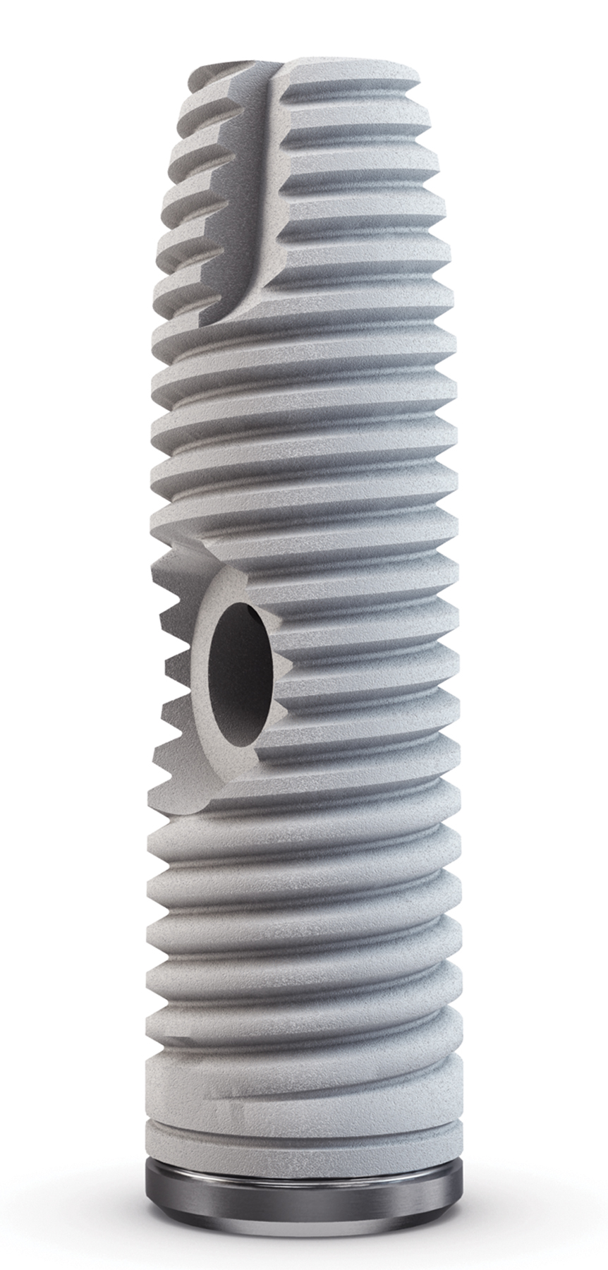
The implant design includes an L form internal channel leading to the apical portion of the implant (Fig. 2), which allows injecting saline and bone graft to the sinus cavity.
FIGURE 2. I – Raise implant with its internal channel

Mechanical test of the implant was performed and compared to Nobel Biocare standard implant. No difference in physical strength was found.
A Sterile NaCl 0.9% is injected through the implant internal channel in order to detach the Schneiderian membrane from the sinus floor.
Aspiration back of the saline is followed by injection of gel form bone grafting material through the same implant internal channel thus filling the space between the sinus floor and the membrane.
The last step is to insert the full implant to the augmented bone.
The hydraulic lift of the sinus membrane is done through the alveolar crest. The minimum bone height required for this crestal approach is 3 mm. Once the implant is inserted completely, the internal channel is closed in the bone without communication with the implant prosthetic platform, preventing penetration of bacteria from the oral cavity to the bone grafted area following the implant insertion.
Case Presentation:
A 40-year-old, healthy woman presented to the dental office. Clinical and radiographic examination revealed: Missing tooth #15. Residual alveolar ridge height was at 5 mm. The treatment plan included insertion of an endosseous implant followed by an implant-retained crown.
In order to be able to apply this plan, a sinus augmentation was required. The I Raise implant was used in this case allowing placing the implant and hydraulic elevation of the sinus membrane simultaneously.
Prior to the surgery, 1000 mg of amoxicillin was prescribed as a prophylactic treatment. A full thickness mucoperiosteal flap was performed. The exact point of implant insertion was marked in the area of tooth #15. Special drills were used to engage the cortical bone of the sinus floor (Fig 3). A diamond bur was then used to cross the cortex bone. The use of a diamond bur will prevent rupture of the Schneiderian membrane (Fig. 4).
FIGURE 3. I – Raise special flat drill
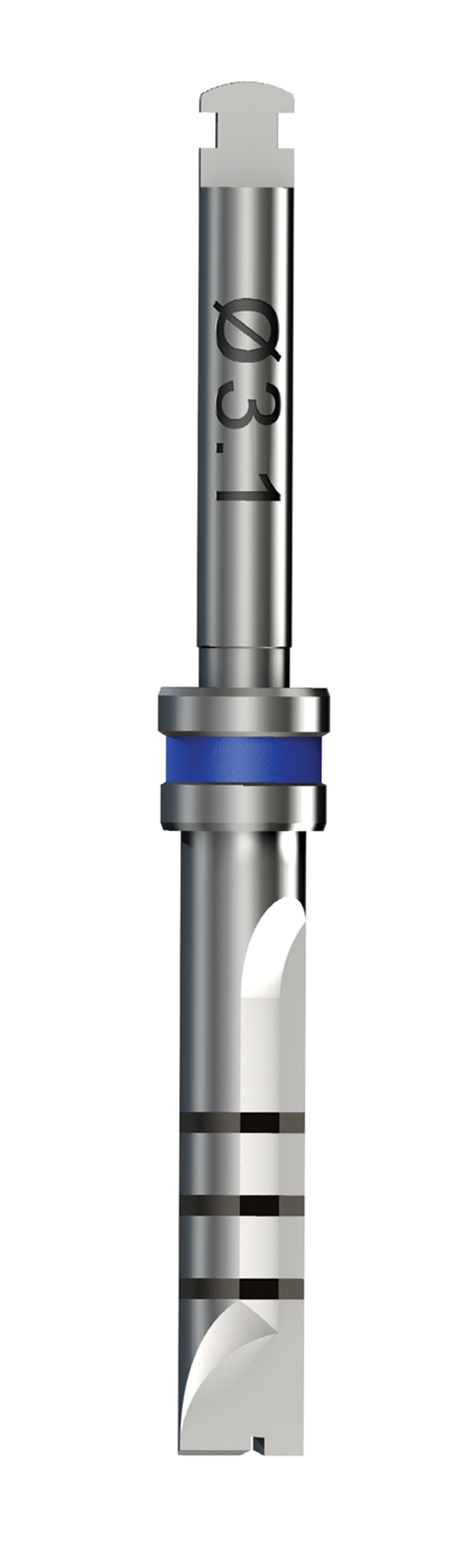
FIGURE 4. I –Raise diamond drill

An I Raise implant of 4.2 mm diameter and 14.5 mm length was inserted half-way. The orifice of the internal channel reached the bone and was placed facing the buccal side. The implant connector was attached to the implant orifice.
Next, 2 ml NaCl saline was injected through the connector in order to detach the sinus membrane by hydraulic equal pressure. Valsalva maneuver-test was performed to confirm membrane integrity.
Aspiration of the saline was followed and a mixture of saline with blood appeared in the syringe, indicating that the Schneiderian membrane was detached and elevated and the blood capillaries were ruptured.
The following stage was the injection of 2 ml synthetic bone grafting material type (MBCP Gel manufactured by “Biomatlante”) TCP/HA in a gel form. The connector was removed and the implant was inserted to its full length, at crest level.
FIGURE 5. Patient Panoramic X-ray prior to the treatment

FIGURE 6. Post treatment Panoramic X-ray
A Cone Beam C.T. (CBCT) was done immediately post treatment, demonstrating a beautiful four-layer creation: air, water, bone graft material, and residual alveolar ridge. At the same time, we noticed the integrity of the Schneiderian membrane together with a healthy sinus.
The I Raise implant internal channel could be seen closed by the injected bone. The surgery ended by closure of the flap followed by conventional sutures. The patient easily tolerated the surgery and immediately returned to her routine life. No side effects, such as swelling, pain or hematoma were reported.
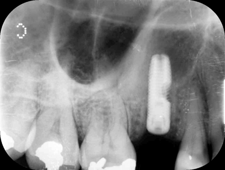
A follow-up at three and six months post surgery were done and the enclosed peri apical X-rays showed calcification, which leads to bone formation (Figs. 7 & 8).
FIGURE 7. Three-month follow up

FIGURE 8. Six-month follow up

Discussion:
I Raise sinus lift technique is friendly and an easy-to-perform surgery. Two separate surgeries are fused to one short surgery to create a minimally invasive procedure, which is well tolerated by the patient, and allows a quick return to normal life, as opposed to other sinus lift surgery approaches. The open lateral window technique, for example, causes substantial side effects such as swelling, pain, hematoma, and long-term recovery. The present hydraulic minimally invasive sinus lift technique is a routine procedure done in private practices and in hospitals. OH
Mili Harel-Raviv, DMD, Former head department, Faculty of Dentistry, McGill University, Montreal, Canada. Director of Raviv Implant cadaver courses worldwide (www.ravivdentalcourses.com).
Oral Health welcomes this original article.
References:
1. Tatum H Jr. Maxillary and sinus implant reconstructions. Dent Clin North Am; 30:207-29.1983
2. Raghoebar GM, Batenburg RH, Timmenga NM, Vissink A, Reintsema H. Morbidity and complications of bone grafting of the floor of the maxillary sinus for the placement of endosseous implants. Mund Kiefer Gesichtschir. 1999;3 Suppl 1:S65-9.
3. Katranji A, Fotek P, Wang HL. Sinus augmentation complications: etiology and treatment. Implant Dent. 2008;17:339-49.
4. Schwartz-Arad D, Herzberg R, Dolev E. The prevalence of surgical complications of the sinus graft procedure and their impact on implant survival. J Periodontol. 2004;75:511-
5. Regev E, Smith RA, Perrott DH, Pogrel MA. Maxillary sinus complications related to endosseous implants. Int J Oral Maxillofac Implants. 1995;10:451-61.
6. Mardinger O, Poliakov H, Biteltum I, Nissan J, Chaushu G. Patient’s perception of recovery after sinus floor augmentation – A prospective study. J Periodontol. 2009;80:572-6.
7. Chen L, Cha J. An 8-year retrospective study: 1,100 patients receiving 1,557 implants using the minimally invasive hydraulic sinus condensing technique. J Periodontol. 2005;76:482-91


