
Introduction
The introduction of cone beam computed tomography (CBCT) to dmf (dentomaxillofacial) radiology in the late 1990s transformed the way the profession as a whole analyzes the oral and maxillofacial complex.1 Dental providers, who previously were limited to two-dimensional (2D) images of three dimensional (3D) anatomical structures, now had the ability to visualize the hard tissues of the head and neck in all the desired planes as well as in 3D reconstructions.
Since the introduction of CBCT is relatively recent, many dentists have not been trained to identify the cross-sectional anatomy visualized in CBCT imaging.
In addition, CBCT scans may reveal occult pathology and incidental findings of varying clinical significance located in structures outside a dentist’s conventional area of expertise.
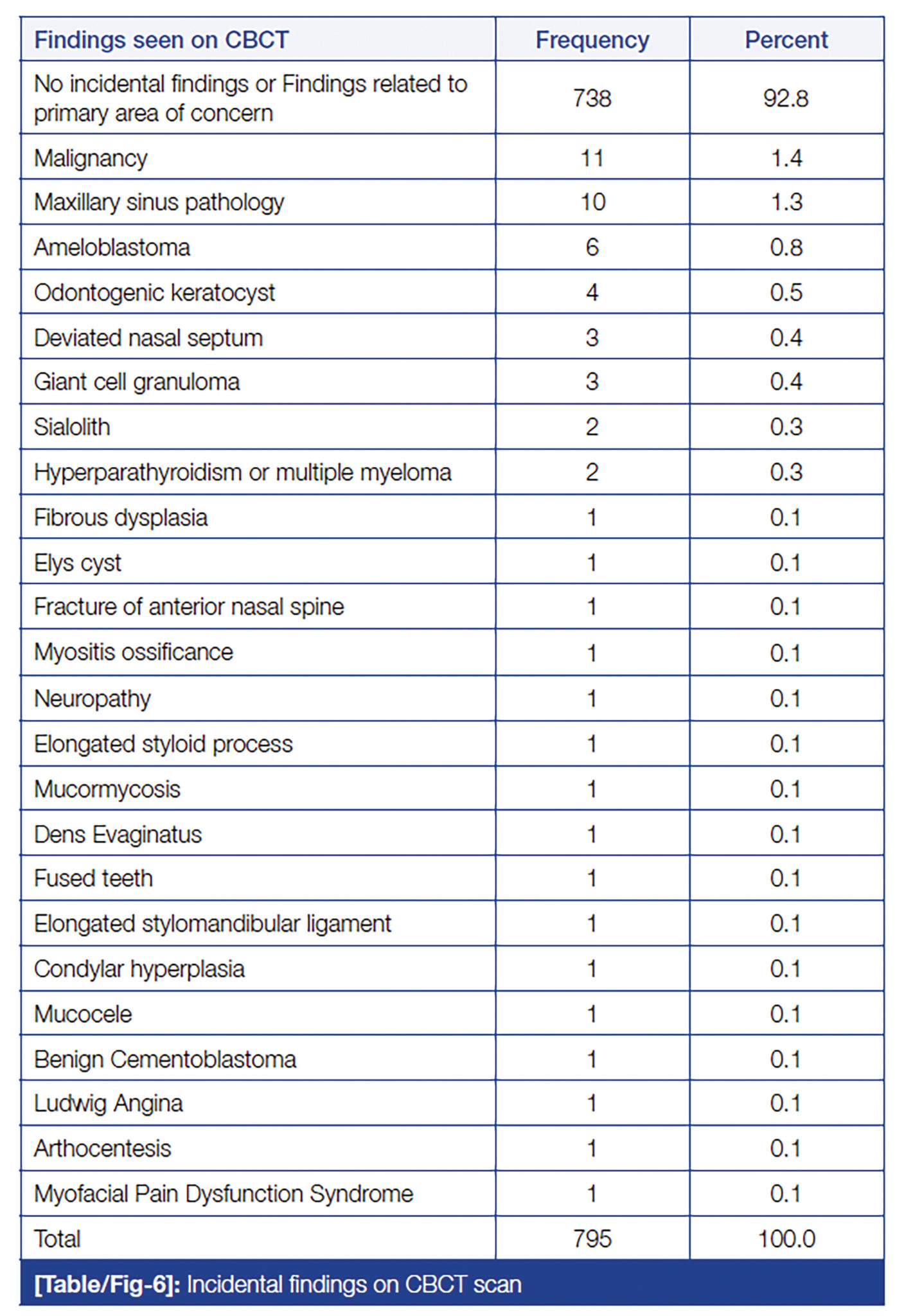
The frequency of incidental findings identified by CBCT has been reported to be as high as 3.2 findings per scan.6 On the other hand, the untrained clinician may have a higher likelihood of reporting false-positive findings, which could result in unnecessary healthcare costs and patient anxiety7 (Fig. 1).
Fig. 1
Dentomaxillofacial radiology is a specialty that is closely related to technology and therefore constantly subject to change.2 The “next big thing” in dmf radiology is not a new type of scanner or storage device or image delivery system, but technologies to improve or accelerate image data interpretation or to facilitate tasks with less or no input from a dentist: this is what most people refer to as “artificial intelligence” (AI) in radiology.8 The recent hype about AI in dmf radiology is mainly due to the success of the tools based on Deep Learning (DL) for analyzing CBCT images. In less than a decade, computers and algorithms based upon DL have gained the power to equal or exceed humans in an increasing number of simple tasks, such as tooth detection and numbering, or the detection of periapical lesions or caries.
Some Definitions
Artificial intelligence is a branch of computer science that describes the research and development of simulated human intelligent behavior in machines. “This involves researching methods that enable a computer to develop intelligent behavior and work independently on problems”.3
Following its inception in 19594 when the first computational trainable neural networks were developed3, the field of medicine and dentistry has witnessed innumerable research using AI (Fig. 2).
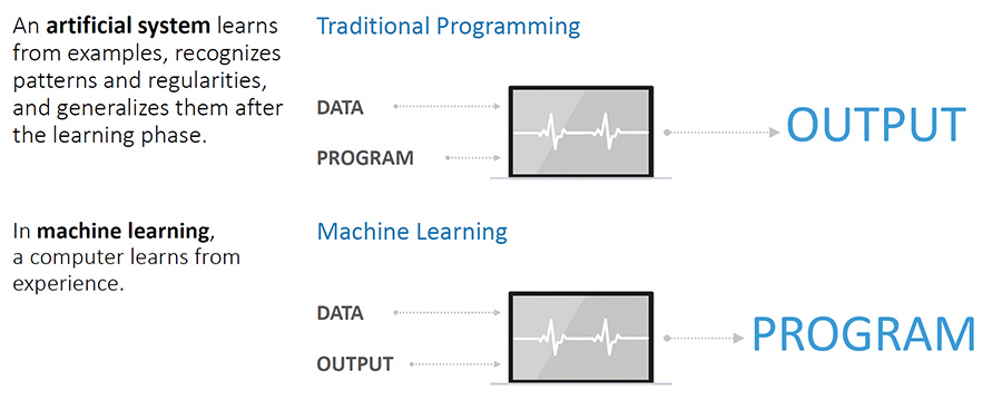
Deep Learning3 is the most widespread machine learning method. It is used by IT companies such as Google, Facebook, and Apple. Computers can autonomously learn from data, such as images.
Deep learning is a class of learnable artificial intelligence (AI) algorithms that allows a computer program to automatically extract and learn important features of input data for further interpretation of previously unseen samples. The key distinction of deep learning methods is that they can learn from a raw data input, e.g. pixels of images, with no handcrafted feature engineering (program) required (Fig. 3).
Fig. 2
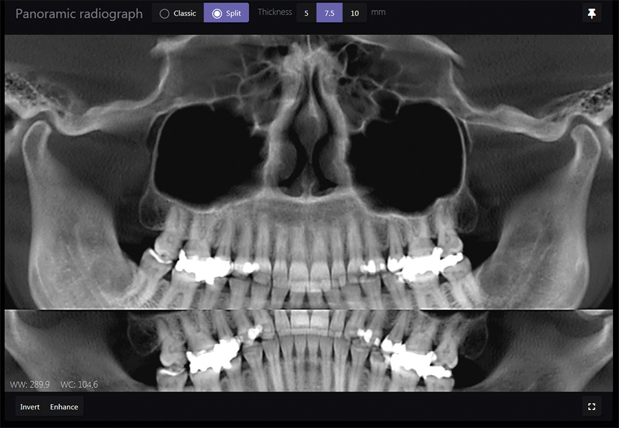
Fig. 3
AI in general5 and AI in dentistry or medicine, in particular, started gaining its foothold with the advent of data computing as well as cloud computing ability and the availability of vast amount of data collected. With the vast amount of data, for example in field of radiology, a specific algorithm can be created which further helps to diagnose and suggest probable treatment options. AI is slowly nudging its head in the field of dental radiology with emphasis on diagnostic records in digital three‑dimensional (3D) scans and cone beam computed tomography. Much information can be gathered and computed to create AI for rapid diagnosis and enhanced treatment planning.
DiagnoCat: AI in Clinical Dentistry
Correct diagnosis is the key to successful clinical practice outcomes. In this regard, adequately trained neural networks like DiagnoCat (Moscow, Russia) can be a boon to diagnosis, especially in conditions having multifactorial etiology. The software proposes a differential diagnosis in its generated description (Fig. 4). This limits the time spent for analysis and provides hints to possible pathological processes, even if those where not primarily indicated.
Fig. 4


DiagnoCat’s artificial intelligence analyzes the acquired CBCT in DICOM format (a standard format in medical imaging) enabling a smooth data transfer. DiagnoCat allows for analysis of CBCT images obtained with any CBCT units without using the installed, unit specific, conventional software (viewer). This increases diagnostic freedom and decreases the addiction to forced software by the manufacturers.
Diagnocat Workflow
A neural network, while processing DICOM files of CT images, finds and segments the main anatomical regions (jaws, teeth, periapical lesions). Diagnocat identifies various conditions and disorders by assessing 50 signs (normal appearance, filling, crown, treated root canal, implant, sign of periapical lesion, etc.) and selects dedicated images to support an individualized therapeutic planning (Fig. 4). The dentist needs a computer connected to the web (desktop, laptop or tablet are all suitable). CT images and reports are stored in the dentist’s personal account. The dentist thus receives a data storage system platform in which data may be arranged by patient name, medical condition, creation and modification dates. CT images may be transferred from dentist to dentist in a protected protocol involving no file transfer procedure.
Clinical Cases Using DiagnoCat
Immediately after uploading files, you obtain access to the Diagnocat Viewer, which automatically produces:
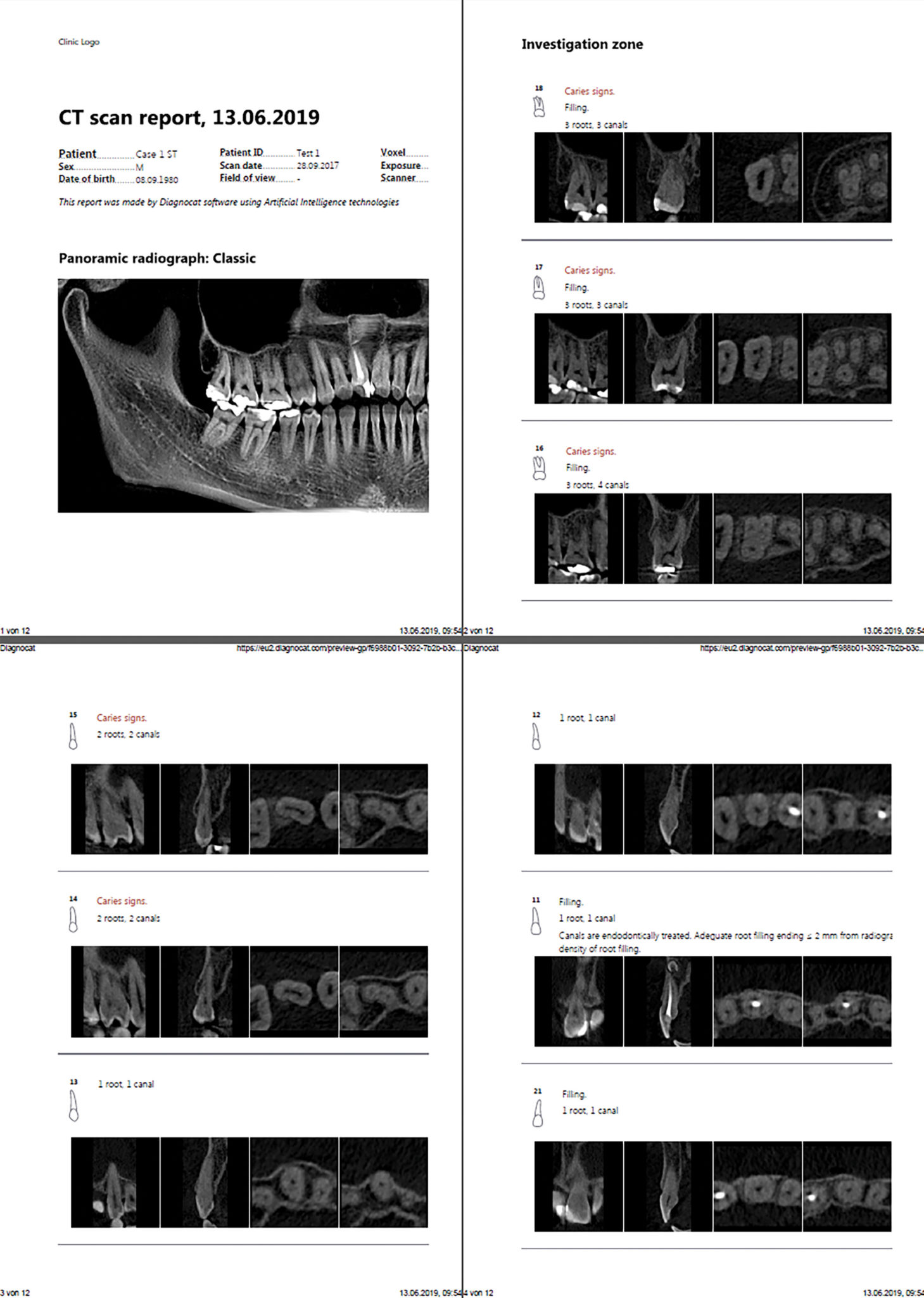
- panoramic view of various thicknesses (Fig. 5)
- a set of slices in three planes for each tooth (Fig. 6)
- a patient report to inform the patient and to motivate him or her to continue and complete the treatment (Fig. 7)
Apart from a panoramic image, the patient report contains the dental chart with annotation in colour on the teeth with findings requiring the patient’s attention marked in red. Other interactive report formats may be generated and obtained from DiagnoCat at the dentist’s request.
Fig. 5
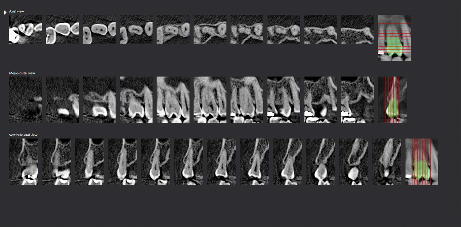
Fig. 6
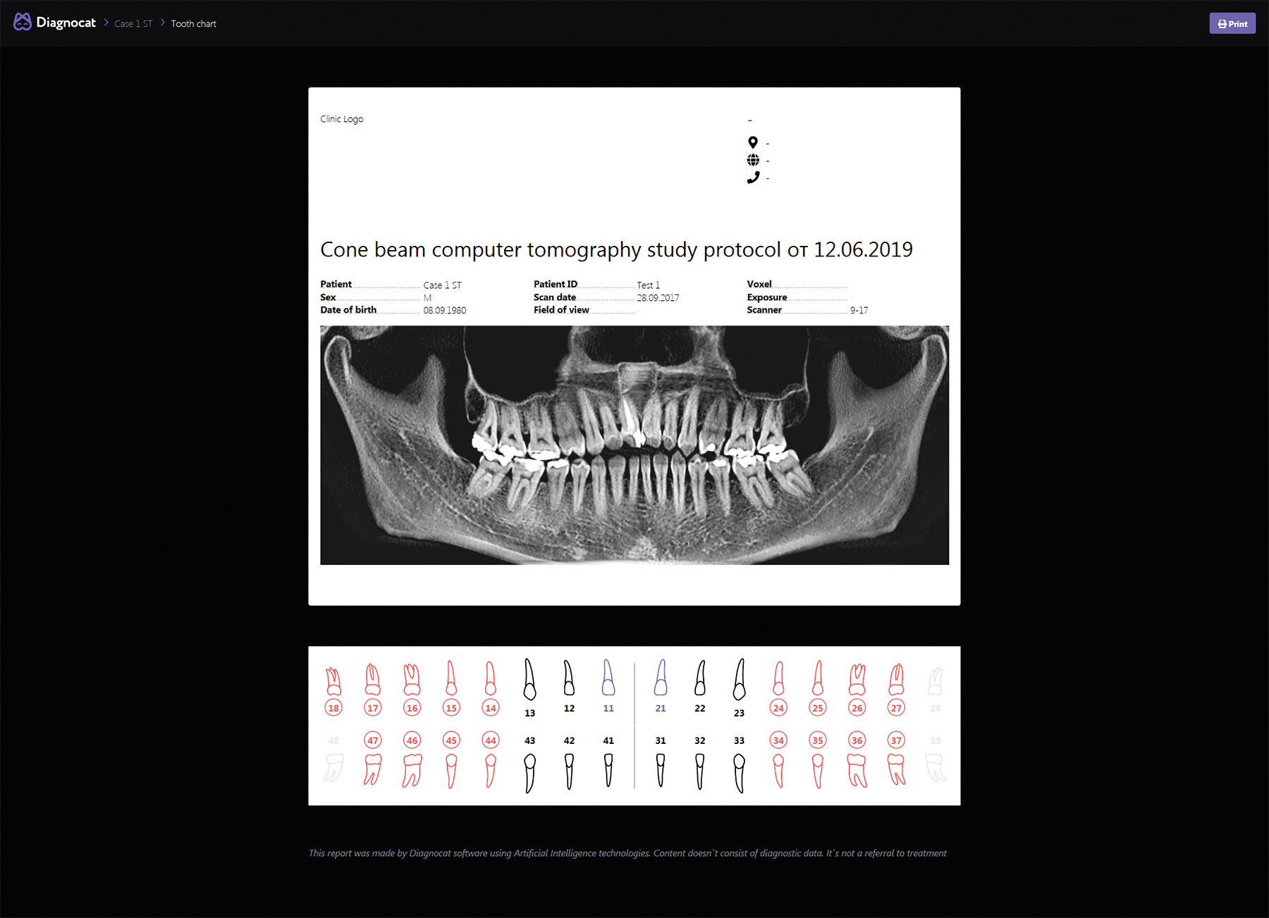
Fig. 7

Clinical Case 1
The most frequent indication for CT is implant planning (Fig. 8). A 53-year-old female patient was referred to our clinic for CBCT based implant planning. The medical history was unremarkable. The radiological evaluation shows no root canal treatments, a dislocated restoration on 34 distal, no root relics or periapical osteolysis, no proof of intrabony lesions. The projected region of implant insertion is without pathological findings. The basal part of the left maxillary sinus presents a cystic alteration. No proof of a focus. When the dataset is uploaded, DiagnoCat automatically generates images of the projected anatomic region that an implant surgeon would need. Depending on the region of interest, DiagnoCat will illuminate the mandibular canal, the bottom of the maxillary sinus, and will calculate measurements between dedicated key points (Fig.9). With this double checked and approved information generated in less than five minutes, the patient was sent back to the referring clinic.
Fig. 8
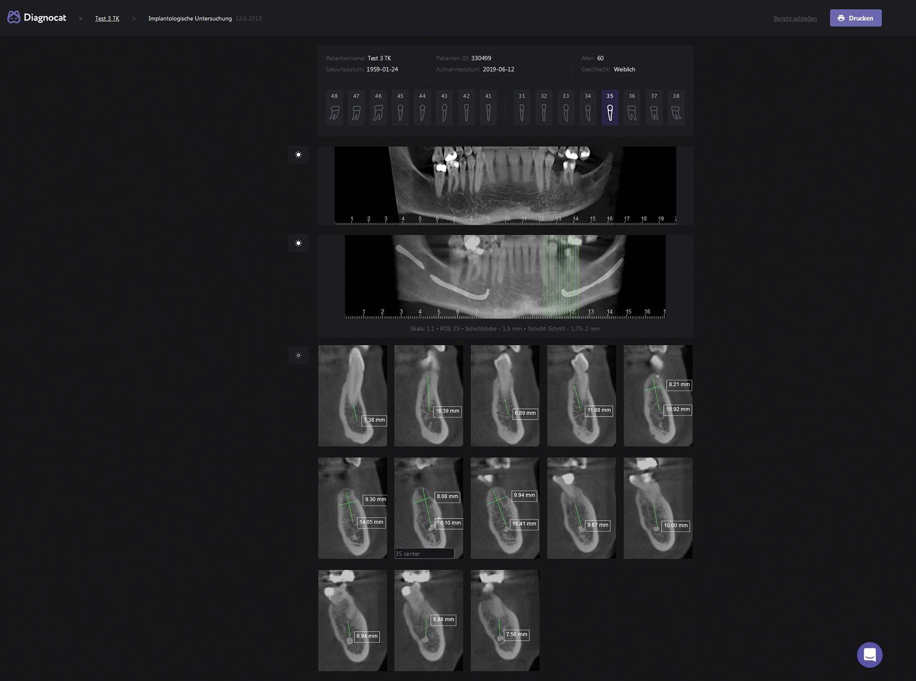
Fig. 9
Clinical Case 2
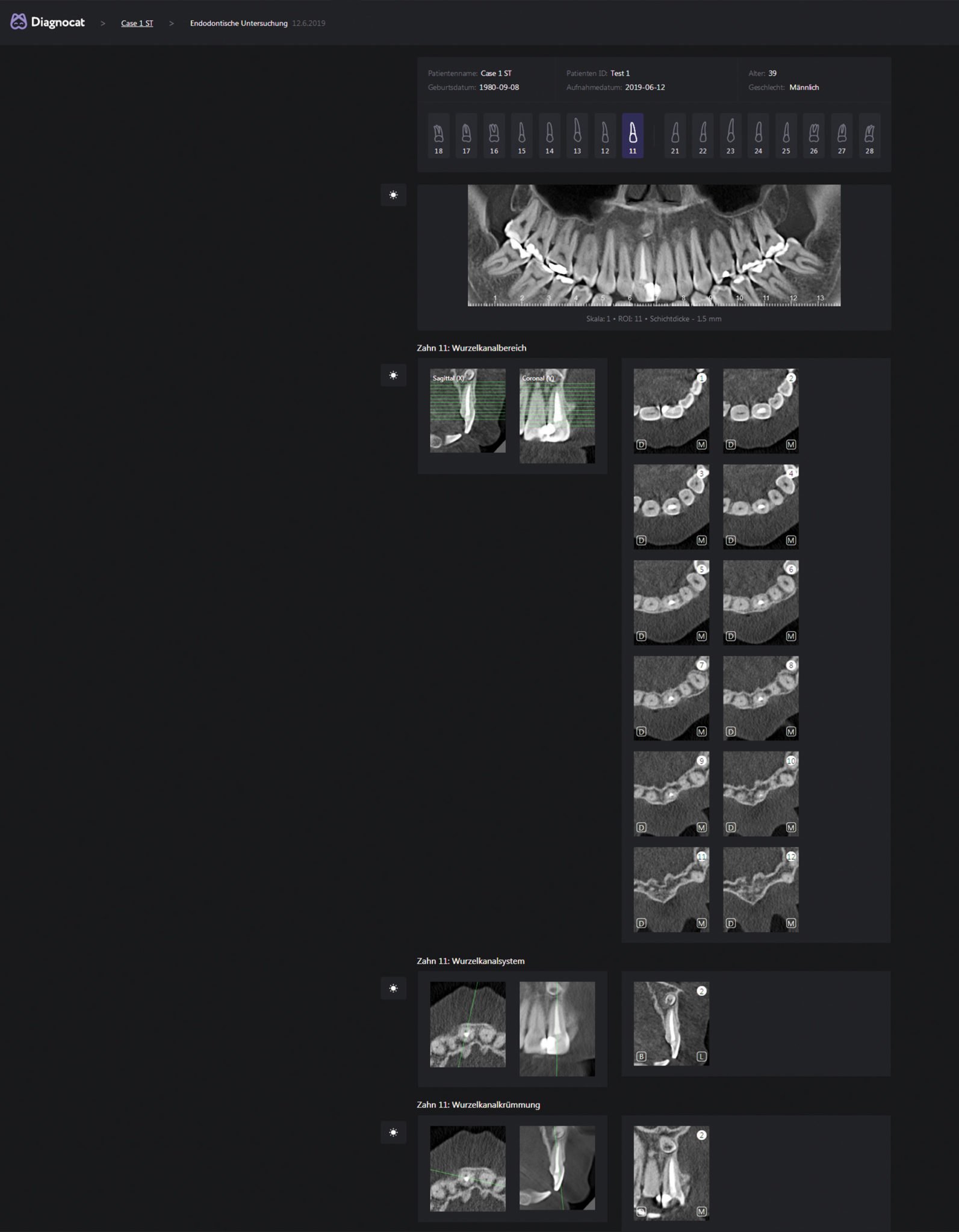
Complicated anatomical structures of root canal systems in endodontics require careful studying of the image. A 32-year-old male patient was referred to our clinic for undefined pain in the maxillary anterior. The medical and dental history was without pathological findings. Uploading the dataset to DiagnoCat, defining the region of interest (ROI), by selecting tooth #11, all images (roots, canals, periapical region) were visualized accordingly (Fig. 10). A hyperdense tooth like structure could be visualized apical of tooth 11. The diagnosis of supernumerary tooth (mesiodens) could be approved. The patient received the medical report and was sent back to the referring clinic for further treatment.
Fig. 10
Report and Documentation Using DiagnoCat
Creating a diagnostic protocol, the documentation of the radiologic examinations, selecting and adapting the dedicated slices of the acquired data (CBCT) requires a lot of time and experience in image editing. DiagnoCat enables recording of diagnostic protocols with just a few clicks. Medical record entries will always be correct, as the DiagnoCat terminology has been carefully examined by an experienced team of dentomaxillofacial radiologists. Supporting images can be added in only a few seconds (Fig. 11).
Conclusion and Preview
Artificial intelligence (AI) is a breakthrough in the field of technology, which is rapidly progressing. Ever since its inception, dentistry has witnessed some of its exceptional achievements. Dentists should get acquainted with this technology as the future of dentistry will surely include implementation of its many applications. DiagnoCat diagnostic solutions enlists the computer- generated knowledge of experts for automated dental radiograph analysis. Computer-aided teeth detection and numbering simplifies the process of filling out digital dental charts. Automation can help save time and improve the completeness of electronic dental records.
While AI cannot replace the role of the dentist or radiologist, accurate and fast processing of CT images by the artificial intelligence of a neural network offers exciting diagnostic opportunities for the future and will certainly be an integral part in point of care dental radiology.
Oral Health welcomes this original article.
References
- Detection of Incidental Findings in Cone Beam Computed Tomography Imaging and Their Clinical Implications, Erika Benavides, DDS, PhD, and Paul C. Edwards, MSc, DDS, FRCD(C), 10 October 2014, https://doi.org/10.1002/9781118674888.ch10.
- Ranschaert, E. Artificial Intelligence in Radiology: Hype or Hope? Journal of the Belgian Society of Radiology. 2018; 102(S1): 20, 1–2.
- Artificial Intelligence Presentation • January 2019 DOI: 10.13140/RG.2.2.28657.74089 Hans-Dieter Wehle, IBM.
- Journal of Medicine, Radiology, Pathology & Surgery • Vol. 5:2 • Mar-Apr 2018 Artificial intelligence: A dentist’s perspective, Anupama Kalappanavar, S. Sneha, Rajeshwari G. Annigeri, Department of Oral Medicine and Radiology, College of Dental Sciences, Davangere, Karnataka, India.
- Sonali Vijay Deshmukh, Editor‑ in‑Chief, Journal of International Clinical Dental Research Organisation, Department of Orthodontics, Dr. D. Y. Patil Dental College and Hospital, Dr. D. Y. Patil Vidyapeeth, Pune, Maharashtra, India, 2018 Journal of the International Clinical Dental Research Organization Published by Wolters Kluwer–Medknow.
- Price JB, Thaw KL, Tyndall DA, Ludlow JB, Padilla RJ. Incidental findings from cone beam computed tomography of the maxillofacial region: a descriptive retrospective study. Clinical Oral Implants Research. 2012; 23(11):1261–1268.
- Kapila et al, Current status of cone beam computed tomography imaging in orthodontics, January 2011Dentomaxillofacial Radiology 40(1):24-34, DOI: 10.1259/dmfr/12615645; Ahmed et al., 2012, Application of cone beam computed tomography in oral and maxillofacial surgery, M Ahmad, J Jenny, M Downie, First published: 29 February 2012 https://doi.org/10.1111/j.1834-7819.2011.01661.x.
- King, BF. Artificial Intelligence and Radiology: What Will the Future Hold? JACR. 2018 Jan 18; 15(3):1–3.
About the Author
 Jörg Mudrak, received his journeyman`s certificate as a dental technician in 1987, started his dental education in 1988 at the FU Berlin and graduated in1993 from the JLU University, Giessen, Germany. He passed his postgraduate specialization for oral surgery in 1997 and works part time in a private clinic for oral surgery. Jörg works globally as a clinical consultant for several companies, focusing on education and scientific research in the area of DMF/ CBCT radiology. Today his main area of interest is in data analysis and artificial intelligence in dental radiology.


