
Introduction
In the academic year of 2014 to 2015, a new continuing education program, Residency in Cosmetic Dentistry, was introduced to the dental community. This program was meant to provide opportunity for practicing dentists who are interested in enhancing their knowledge and skill in the subject of esthetic dentistry to join. The program included teaching instructions through lectures, small group seminars as well as hands-on clinical experience. The lectures were provided by a group of carefully selected speakers and covered a wide range of topics. Clinical procedures that were performed by participants ranged from ceramic veneers and crowns, CAD-CAM and implant-supported crowns and direct resin composite restorations. The aim of this paper is to share one interesting case treated by one of the program participants (KM) with readers of Oral Health.
Following the discovery of acid etching and bonding to enamel, the porcelain veneer technique was first introduced to the profession in the 1980s by Dr. John Calamia1. Its swift adoption and vast utilization in dentistry made it a standard dental procedure over a short period of time. However, long-term success and longevity of porcelain veneers continues to necessitate careful case selection.2,3 Compared to crowns, veneers offer a conservative preparation design with minimal depth, ranging from .3 to .5 mm, terminating in enamel that is necessary for reliable bonding.2 In a long-term clinical study that evaluated longevity of 580 porcelain veneers over a period of 12 years, when preparations had 20 percent involvement of dentin failures were observed, however, when preparations were completely confined to enamel no veneer failures were observed.4 This finding was confirmed in a review article on the topic that analyzed 24 papers published on the survival of porcelain veneers.5 It stated that “there is reasonable evidence indicating that a veneer preparation into dentin adversely affects survival.” When preparing teeth to receive porcelain veneers dentists must endeavor to maintain preparations minimally invasive with no involvement of dentin where possible.
While original porcelain veneers were made using feldspathic porcelain formulations, new glass-ceramic formulations with superior strength and resistance to chipping have been developed over the years and are now being utilized in place of the original formulations.
According to Nohl et al., “A complete understanding of a patient’s aesthetic problems is the key to treatment planning. Only then can an attempt be made to match expectations with realities and to provide appropriate restorations”.6 In the case presented below a strategic and instrumental approach was adopted throughout in order to ensure that the outcome met the patient’s expectation and at the same time the most conservative and most effective treatment approach was followed.
Case Report
A 44-year-old female with a history of bulimia presented with dark discoloration in her upper anterior teeth (Figs. 1-3). Vital bleaching of the anterior teeth was attempted, however, while there was some improvement in color the outcome remained less than ideal. Therefore, the case was assessed for suitability for restoration with ceramic veneers. A group discussion of the case among residency program participants and instructors ensued and the case was deemed suitable for restoration with glass-ceramic veneers. A diagnostic wax-up was made in the laboratory for assessment of suitability of the veneer restorations and case discussion with the patient. The patient was shown the diagnostic wax-up and the procedure for preparation of the teeth was explained to her in full detail. Alternative treatment modalities with their pros and cons were also discussed with the patient. The final treatment plan that was formulated with the patient’s consent and approval included six glass-ceramic veneers for maxillary anterior teeth, glass-ceramic veneers for maxillary first premolars and one glass-ceramic crown for maxillary second premolar and a glass-ceramic veneer for the other.
FIGURE 1. Preoperative appearance of the maxillary anterior teeth. Several bleaching attempts were made to get rid of the underlying dark discoloration with little response.

FIGURE 2. Preoperative appearance of the right anterior side.
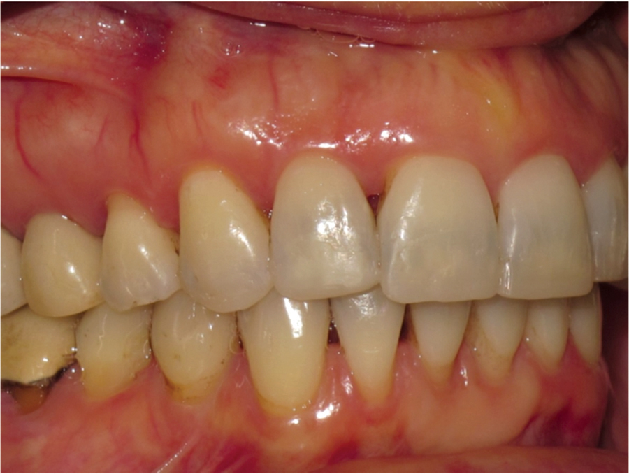
FIGURE 3. Preoperative appearance of the left anterior side.

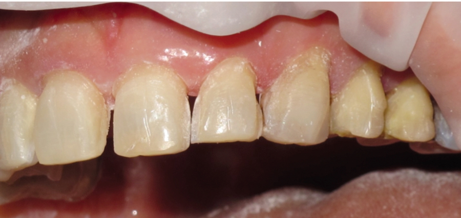
Teeth were prepared for veneers with minimal tooth reduction that terminated in enamel using special diamond burs (#M392-016 and #SF379-023, Axis Dental) (Fig. 4). For premolar veneer preparations a small shelf was made with bur # F856-016 (Axis Dental) on the buccal aspect for better stability during function. The second premolar to receive crown was prepared with a parallel-sided flat-end diamond bur with a 1 mm all-around shoulder finish line and occlusal reduction of 1.5 mm. All line angles were maintained rounded to minimize stress build-up during function (Fig. 4). The diagnostic wax-up was also used to fabricate a reduction guide used to ensure adequate reduction prior to impression taking. A custom made perforated acrylic resin tray was used for impression taking with a polyvinyl siloxane material (Aquasil light- and heavy-body, Dentsply).
FIGURE 4. Appearance of prepared anterior and premolar teeth after removal of provisional restorations. Both 1st and 2nd premolars were prepared with a step on the vestibular aspect to help in stability of the porcelain veneers.
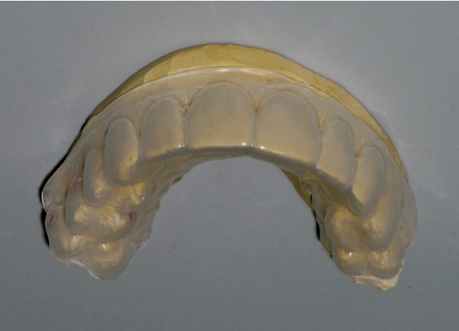
The diagnostic wax-up was also used to fabricate a matrix for making provisional restorations (Fig. 5). Spot-etching of enamel with phosphoric acid gel for 30 seconds was performed at mid-central locations on facial aspects of canines and premolars. Bonding resin was applied on the etched spots and light-polymerized. The matrix was loaded with provisional veneer material (Luxatemp, DMG America) and secured in place over the prepared teeth. After three minutes the matrix was then lifted off with a sickle scaler and trimming of excess material was carried out with a #7901 tungsten carbide bur. Occlusion was checked and adjusted accordingly. Final polishing with polishing discs and points was then performed to a glaze-like finish (Fig. 6). A provisional crown was made for the second premolar that was prepared for a crown using the same material.
FIGURE 5. A clear matrix was made in a vacuum-forming machine on the waxed up model for fabrication of provisional veneers.
One crown and nine glass-ceramic veneers were fabricated at a local dental laboratory (Shaw labs) in lithium disilicate (e.max CAD, Ivoclar-vivadent) (Figs. 6-9). At the next appointment the provisional restorations were carefully removed and teeth surfaces were cleaned with a slurry of medium-grit pumice with a rubber cup in a slow-speed handpiece. Intaglio surfaces of the ten glass-ceramic restorations were first etched with hydrofluorice acid gel then silanated with a silane-coupling agent. All ten restorations were carefully tried-in while the patient remained in a near horizontal position on the dental chair with face up. The patient was shown the restorations in a mirror for approval. Cementation of the nine glass-ceramic veneers followed using a light-polymerized resin cement (Variolink II, Ivoclar-Vivadent). First enamel surfaces of the prepared teeth were etched with phosphoric acid-etching gel for 45 seconds. The two veneers for the centrals were first simultaneously cemented (Fig. 10). After seating the veneers excess cement was carefully removed with an explorer before light-polymerization through the veneer material (Blue-phase Style, LED light-curing unit, Ivoclar-vivadent). This was followed by cementation of the maxillary laterals’ veneers
FIGURE 6. Provisional restorations were made using a clear matrix made to a waxed-up model. Spot bonding at the centers of the vestibular surfaces of the premolars was followed to optimize retention.
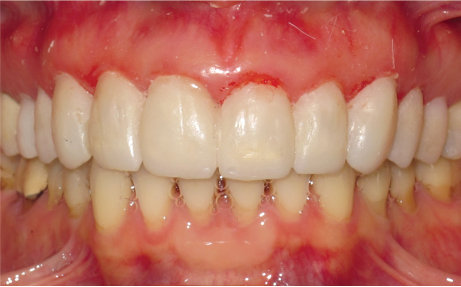
FIGURE 7. Nine porcelain veneers and one porcelain crown were fabricated at a dental laboratory.
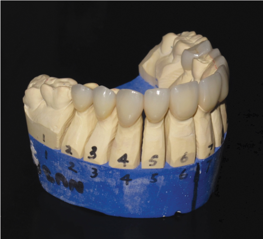
FIGURE 8. Lingual aspect of the fabricated porcelain restorations.

FIGURE 9. Four porcelain veneers and one crown fabricated for the right side.
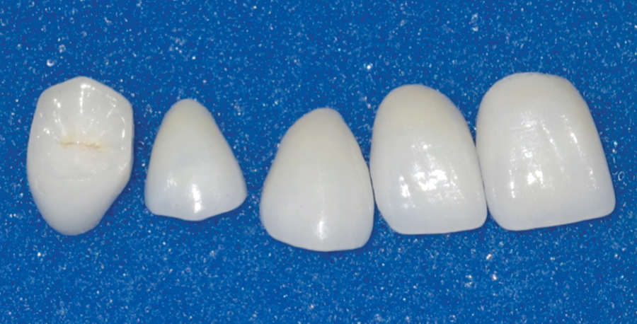
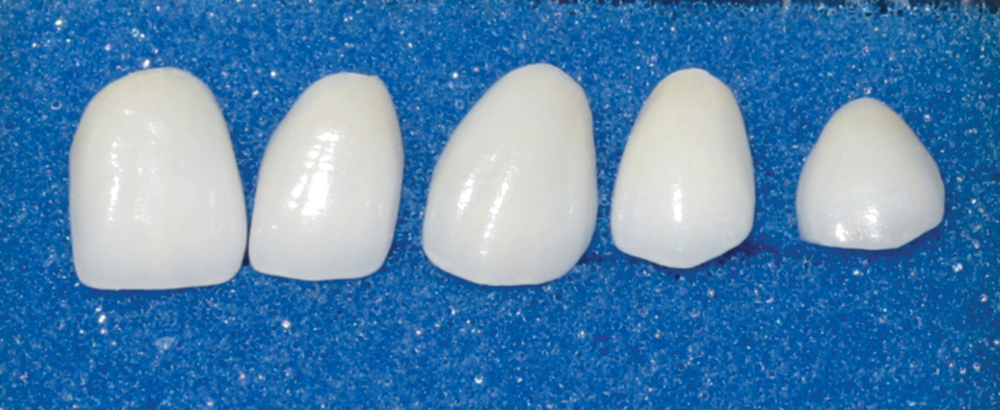
FIGURE 10. Five porcelain veneers fabricated for the left side.
(Fig. 11), then veneers for maxillary canines and first premolars were cemented one-side at a time (Figs. 12-14). Finally, the last veneer and the glass-ceramic crown were cemented to the maxillary second premolars. Variolink II was used in the dual-cure mode for cementation of the crown. Figure 15 shows a post-operative view of the case after excess cement was removed and interproximal areas thoroughly polished.
FIGURE 11. Porcelain veneers of the maxillary central incisors were first cemented in place with a light-polymerized resin cement.
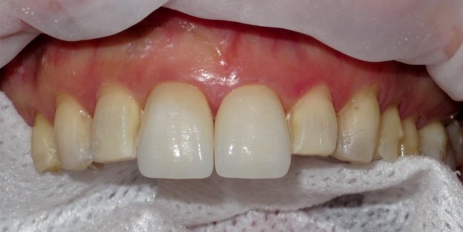
FIGURE 12. Veneers for the laterals were then cemented.
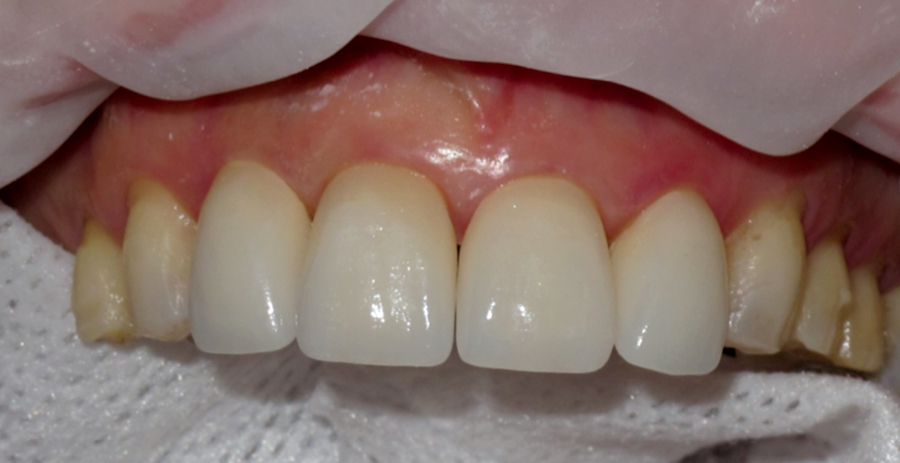
FIGURE 13. Veneers for canines and first premolars are cemented.

FIGURE 14. Right side view showing porcelain veneers on central, lateral, canine and first premolar in place.
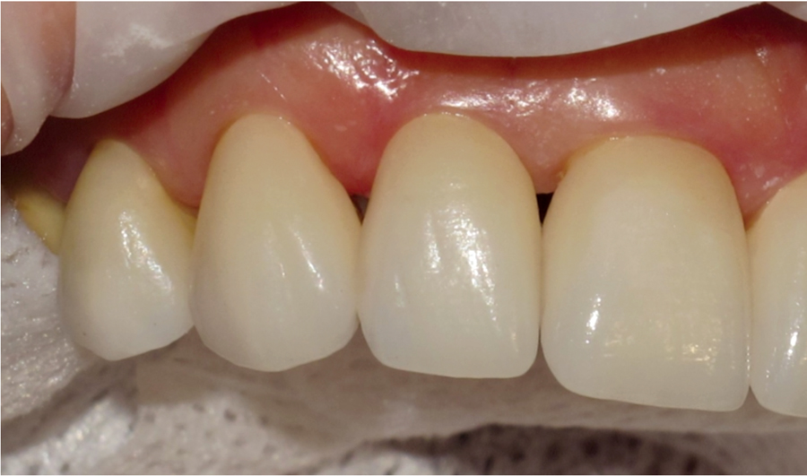
FIGURE 15. Left side view showing porcelain veneers on central, lateral, canine and first premolar in place.
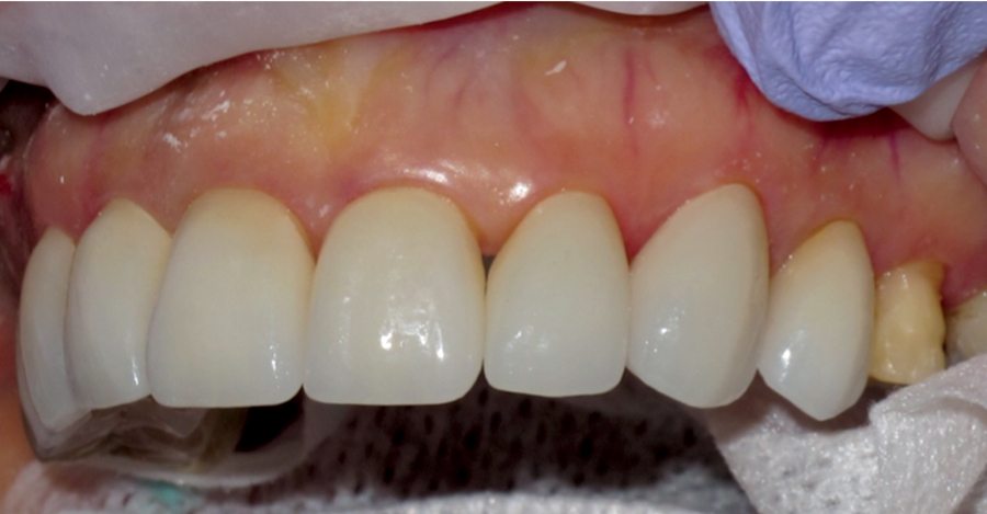
FIGURE 16. Frontal view showing all nine porcelain veneers cemented in place in addition to the porcelain crown cemented on tooth 15.

Conclusions
When considering treatment of maxillary teeth with veneers to improve esthetics dentists must consider each and every case according to its own merits. While the tooth preparation procedure is fairly simple great care must be taken to ensure that preparation boundaries terminate in enamel without encroachment into dentin. Cementation of multiple veneers can be a little tedious, however, breaking them down into manageable numbers is recommended. Careful removal of excess cement after setting is important to maintain health of the gingival tissues. When used judiciously in carefully-selected cases ceramic veneers can help patients achieve the most desirable esthetic appearance. OH
Dr. Omar El-Mowafy is tenured professor and head of restorative dentistry at the Faculty of Dentistry, University of Toronto. He is a full-member of the School of Graduate Studies of the University of Toronto. Dr. El-Mowafy is also member of Omicron Kappa Upsilon (Honor Dental Society). He published over two hundred research papers, case reports, literature reviews, book reviews in peer-reviewed journals with high scientific impact factor. He is member of Editorial Boards of Operative Dentistry and International Journal of Prosthodontics. He also, acts as reviewer for over 12 international journals. Dr. El-Mowafy spoke at scientific meetings at 29 different cities worldwide in addition to speaking on-board of two cruise ships. He maintains position in private practice in Mississauga since 1989.
Oral Health welcomes this original article.
References
1. Calamia JR. Etched porcelain veneers: the current state of the art. Quintessence Int 1985; 16(1):5–12.
2. El-Mowafy OM. The use of both porcelain veneers and all-porcelain crowns in restoring anterior teeth. J Can Dent Assoc 2006; 72(9):803–6.
3. El-Badrawy WA, El-Mowafy O. Porcelain veneers Vs. crowns when resolving esthetic problems – two case reports. J Can Dent Assoc. 2009;75(10):701-4.
4. Gurel G, Morimoto S, Calamita MA, Coachman C, Sesma N. Clinical performance of porcelain laminate veneers: outcomes of the aesthetic pre-evaluative temporary (APT) technique. Int J Periodontics Restorative Dent. 2012, 32(6):625-35.
5. Burke FJ Survival rates for porcelain laminate veneers with special reference to the effect of preparation in dentin: a literature review. J Esthet Restor Dent. 2012;24(4):257-65.
6. Nohl FSA, Steele JG, Wassell RW. Crowns and other extra-coronal restorations: Aesthetic control British Dental Journal 2002, 192, 443 – 450.


