
In years past, traditional restorative care relied on treating damaged and decayed teeth with aggressive preparation to place a strong, firm restoration. The emphasis was on the strength of the restoration, and less about the function and biomechanics of the final restored tooth. Unfortunately, this often led to weaker teeth and complications such as root canals with treatment becoming progressively more aggressive and invasive.
Biomimetic dentistry is intended to copy what is life-like, preserving the ideal properties of natural teeth while using minimally invasive preparations to conserve enamel and dentin. This is done with a better understanding of the caries process and carefully placed bonded materials to replace lost tooth structure and regain strength and beauty.
Fortunately, we now have bioactive restorative materials that are able to promote tooth remineralization and/or inhibit tooth demineralization.1 They help regain tooth structure by creating an optimal pH, bacterial inhibition, biofriendly tissue tolerance, tooth like physical properties, seal from noxious intrusion, and long-term stability.2,3
A newer product category called “giomer” has been proven in many research studies to overcome the weakness of compomers which are often technique sensitive, have limited fluoride release, and are affected by water sorption which decreases physical properties with time. Giomer is a tooth-colored restorative material that uses a resin base and pre-reacted glass ionomer (PRG) technology.
S-PRG technology delivers some properties of glass ionomer such as fluoride release and recharge which helps prevent caries recurrence. It also provides properties of composite resin such as excellent aesthetics, easy polishability, biocompatibility and smooth surface finish. Moreover, it has an antiplaque effect by forming a material film layer with saliva that is reported to minimize plaque adhesion and inhibit bacterial colonization.4 Giomers report comparable microleakage scores to that of composite resins in permanent teeth.5,6
“Giomer restorative material showed lower microleakage scores than compomers. Giomer restorative material could be considered a suitable class II restoration of primary molar in high caries risk children.”4
Today’s dental professionals are armed with the tools to predictably restore teeth to biomimetic function combining: the improvements in restorative materials, the enhanced knowledge of the biochemical causes and risk assessment of caries, decay removal using better magnification, caries detection dyes, lasers and a better understanding of crack propagation from occlusion and the biomechanical effects of wider isthmus sizes.
This article is intended to demonstrate the use of Shofu’s innovative giomer technology while using conservative tooth preservation techniques.
Case Presentations
Case #1
A 32-year-old woman had lived out of the country for ten years with no professional dental care. She had a history of high caries risk that had been resolved and had fallen into some bad habits while travelling. Her complete examination demonstrated class 5 lesions in the molar areas due to poor home care (Fig. 1). Since she had no areas of cold sensitivity a vigorous hygiene and remineralization protocol with CariFree products (CariFree, Albany OR) was initiated, prior to restorative care with Beautifil II (Shofu, San Marcos CA).
After isolation with the Isolite system (Isolite, Goleta CA) decay was removed using a Lite Touch Erbium YAG laser (AMD Lasers, Indianapolis IN) at settings 1Milijoule and 20 Hz with no anesthesia (Fig. 2). A 2 mm bevel was placed coronally to blend the future composite into the enamel for biomechanical and esthetic reasons. Enamel and dentinal micro-enhancement was performed using light air abrasion with 27micron Aluminum oxide at 40 psi then rinsing with Chlorohexidine and water. Then the enamel was selectively etched with 37% phosphoric acid, thoroughly washed with water and gently dried to leave the surface moist and not desiccated (Fig. 3).
BeautiBond (Shofu) was applied in one coat with a microbrush, was let stand for 10 seconds without agitation, then gently air-dried for three seconds to dry and spread the material. Finally, it was light cured for 5 seconds with an LED light (10 seconds with a halogen is also sufficient) (Fig. 4). Beautifil Flow Plus was applied to the dentin and cured for 20 seconds (Fig. 5). The higher viscosity Beautifil II (Shofu) was placed using the convenient compule system and sculpted with a gold Microfil composite instrument (Almore, Beaverton OR) then feathered towards the margin using a small artist brush and sculpting resin and finally light cured for 20 seconds (Fig. 6). After placing topical anesthetic, finishing and polishing were performed using a 12 bladed EF 9 carbide bur (Microcopy, Kennesaw GA) and Shofu’s One Gloss pointed polisher (Figs. 7 & 8). The patient reported no sensitivity post operatively (Fig. 9).
Figure 1

Cervical decay around molars was due to non-optimal plaque control and lowered salivary pH.
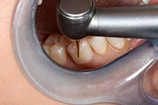
Figure 2

Removal of decay is predictably performed with no anesthesia using an Erbium YAG laser, followed by placing a long superior bevel with a flame shaped diamond and microabrasion using 27 micron aluminum oxide at 40 psi to improve the seal.
Figure 3

Selective etching the enamel with 37% phosphoric acid for 15 seconds is followed by gentle water rinse and air-drying without desiccation.
Figure 4

Shofu’s BeautiBond is applied in one coat with a microbrush with no agitation needed.
Figure 5

Beautifil Flow Plus is placed as dentinal replacement.
Figure 6

Beautifil Flow Plus is placed as dentinal replacement.
Figure 7
Initial finishing is performed with an EF 9 composite finishing bur after placing topical anesthetic.
Figure 8

Shofu One Gloss PS aluminum oxide point conveniently polishes the composite surface to minimize stain
and plaque adhesion.
Figure 9
The final restorations create a bioactive and conservative barrier to decay.
Case #2
A healthy 52-year-old nurse presented for a cleaning and exam with no symptoms. Areas of redecay were noted around several restorations. Initial treatment focused on areas of immediate concern. The lower right had some biomechanical risks especially the large amalgam with a wide isthmus and a previous pulp cap. Ideally, a ceramic restoration will be placed in the future. However, given the potential of pulpal consequences, the patient preferred a conservative direct restoration initially (Figs. 10 & 11).
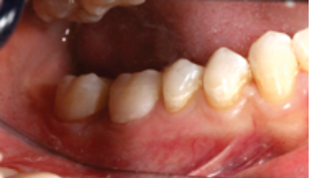
Figure 10

An asymptomatic quadrant of teeth with twenty year-old amalgams that are showing early signs of microleakage and microcracks around the marginal interface.
Figure 11

Preoperative radiographs demonstrate lost dentin from restoring the previous cavities and potential undermining of the marginal ridges. A previous pulp cap was noted on tooth #47.
After balancing the occlusion using a deprogrammer, restorative care was initiated. The bite was reverified prior to anesthesia. Using the Isolite system, all amalgam materials were removed and then Seek (Ultradent, South Jordan UT) was placed to locate areas of infected dentin and cracks (Fig. 12).
The weak enamel and remaining decay were conservatively removed using the Lite Touch (AMD Lasers, Indianapolis, IN) at 150 Milijoules and 20 Hz (Fig. 13) followed by the application a very thin layer of Theracal (Bisco, Schaumburg IL), which was light cured with an LED wand (Figs. 14-16).
Figure 12

After removal of the metal restorations and obvious decay, caries detection dye helps identify remaining areas of decalcification.
Figure 13

An all hard tissue laser helps to conservatively remove remaining debris and preserve a “peripheral rim” of enamel and to conservatively cleanse proximal cracks on teeth #45 and 47 (that did not extend buccolingually). Use of the laser conserves tooth structure.
Figure 14

The enamel and dentin are treated with microabrasion using 27-micron aluminum oxide at 40 psi. Then the preparation is further cleansed with chlorhexidine.
Figure 15
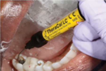
Since deep decay was removed on the pulpal floor of tooth #47, which has been asymptomatic, more invasive procedures were not recommended. The patient was advised of all options and appreciated the conservative approach. Though no pulpal exposure is present, a bioactive layer of TheraCal is applied to the area along the crack to provide an antibacterial high pH barrier to promote remineralization.
Figure 16

Note that TheraCal is placed in a thickness of 1 mm to allow for a full cure and does not affect the strength of the future composite.
Selective etching with phosphoric acid of the occlusal and proximal enamel was done to enhance the final seal of the restoration (Fig. 17). Anatomically correct sectional matrices and rings (Garrison, Spring Lake MI) were placed, then the entire preparation was coated with BeautiBond (Fig. 18).
Next the application of a layer of Beautifil Flow Plus acted as an internal dentinal replacement t well adapted to the pulpal floor, internal walls and proximal margins (Fig. 19). An enamel layer of the more viscous Beautifil II was injected into the preparation and sculpted with composite instruments (Fig. 20).
Shaping and polishing were performed with a one-use 12 bladed carbide football and a Shofu One Gloss point. Because of the excellent adaption of the matrix system no interproximal shaping was necessary and the contacts were outstanding (Fig. 21). Post operatively, the patient was ecstatic with the improved appearance and reported no discomfort (Fig. 22). Radiographically, the restorations were dense with anatomically correct contours (Fig. 23).
Figure 17

A selective etch of the marginal enamel is performed for fifteen seconds with 37% phosphoric acid.
Figure 18

After the Garrison 3D Fusion sectional matrices and rings are placed on each tooth, one coat of Beautibond is applied, allowed to let stand for 10 seconds, gently air-dried for 3 seconds, and light cured for 5 seconds with an LED light.
Figure 19

Beautifil Flow (Shofu) is placed into the deepest areas of the preparation for greater adaptation, acting a dentinal replacement.
Figure 20

Beautifil II high viscosity is injected, adapted, and sculpted as an enamel replacement with a plugger and ball burnisher for excellent adaptation.
Figure 21

The composite is adjusted and smoothed with a new one – use 12 bladed carbide football prior to occlusal verification and final polish with a Shofu One Gloss PS aluminum oxide point.
Figure 22

Final clinical result shows a greatly enhanced esthetic and functional restoration.
Figure 23

Post treatment radiograph shows preservation of tooth structure and dense adaptation of the restorative materials.
Summary
Shofu’s innovative giomer restorative materials have been designed to improve acid resistance and remineralize remaining tooth structure. This provides a durable bioactive clinical result that has been demonstrated in eight-year and 13-year patient studies at the University of Florida.5,6 The evidence is reassuring to the patient and clinician. The layered combination of low and high viscosity beautifil materials provides a well-adapted dense restoration that is strong on its own, and enhances the flexural resistance of teeth to decrease biomechanical risk. Giomer use combined with careful removal of previous restorative materials and conservative decay removal using a hard tissue laser helps to preserve and maintain healthy tooth structure and to achieve restorative longevity. OH
Oral Health welcomes this original article.
References
- Chen L, Shen H, Suh BI. “Bioactive dental restorative materials: a review”. Am J Dent. 2013 Aug; 26(4):219-27.
- Jefferies SR. Bioactive and biomimetic restorative materials: a comprehensive review. Part 1. J Esthet Restor Dent. 2014; 26:14-26.
- Griffin, J. “Regenerative Materials Where They Matter Most”. Dentistry CE Today. Volume 34 No. 4: 1-9.
- Eldesouky et al. “Microleakage of Giomer and Compomer in Primary Teeth”. Alexandria Dental Journal. (2016) Vol.41 Pages: 188-193.
- Gordan, V, Mondragon E, Watson RE, Garvan C, Mjör IA. “A clinical evaluation of a self-etching primer and a giomer restorative material: Results in 8 years. J Am Dent Assoc 2007; 138:621-7.
- Gordan VV, Blaser PK, Watson RE, et al. “A clinical evaluation of a giomer restorative system containing surface prereacted glass ionomer filler: results from a 13-year recall examination. J Am Dent Assoc. 2014;145(10):1036–1043.
 Dr. Hugh Flax is a Past President of the American Academy of Cosmetic Dentistry. He has lectured and authored in Europe, Japan, Canada, and the United States on lasers, smile design, and advanced restorative techniques. Dr. Flax received his DDS degree from Emory University in 1983. While doing a residency he did posterior composite research at LSU Dental School in 1984-1985. He has been a member of the American Academy of Cosmetic Dentistry since 1994 and became Accredited in 1997. He was President of the Atlanta Chapter of the AACD (1996-1998) and founded the Georgia Academy of Cosmetic Dentistry in 2007. Dr. Flax served for two years as Co-Chair of the Conference Advisory Committee for 2003 AACD Scientific Session in Orlando. He is on the Editorial Board of the Journal of Cosmetic Dentistry and Practical Periodontics and Restorative Dentistry. He has been the Chairperson of the AACD Private Education Advisory Council, the 2008 AACD Meeting, the 2010 AACD European Meeting, and was a member of the AACD Board of Directors for over 8 years. Dr. Flax is a member of the AAED, ASDA., ADA, AGD, ALD, AAID, ICOI, Catapult Elite, and the L.D. Pankey Alumni Association. He is a certified Fellow with World Clinical Laser Institute, Fellow of the International Academy of Dentofacial Therapeutics, Master in the ICOI, Associate Fellow of the AAID, Diplomate of the American Board of Cosmetic Dentistry, and a graduate of the Kois Center. Dr Flax practices full time in Atlanta, Georgia focusing on functional appearance-related conditions and advanced laser dentistry.
RELATED ARTICLE: Giomer Varnish for Prolonged Hypersensitivity Relief
Follow the Oral Health Group on Facebook, Instagram, Twitter and LinkedIn for the latest updates on news, clinical articles, practice management and more!


