
In the many years since the 1985 CEREC introduction led by Dr. Werner Mormann, we have seen digital dentistry grow. Digital dentistry allows for the utilization of technology and material advances to provide exceptional dental care, a diversified treatment philosophy, reduced patient visits and increased case acceptance that enhances the economics of a dental office. In our daily lives, we see technology aiding us with examples being the persuasive use of the internet, mobile phones and computing. Unfortunately, we see technologies like CEREC still being used by only a fraction of dentists to a fraction of their potential. Part of this is natural resistance to any new technology adoption curve. Another part of this resistance to positive digital technology is the fear that new ways produce lower quality results than the previous traditional system. In 2016, proper workflow and the adoption of technology, make that argument a reference to a completely different era.
In this article, we hope to help dentists understand the basics of CAD/CAM (CEREC) execution in the modern dental practice. There is a wider point to be made about how technology never really achieves its potential until it is embraced by the team and is fully accepted, but the first step is technical excellence in the use of a digital device to ensure that the quality of clinical outcome is never at issue.
About CEREC
CEREC, by Sirona Dental Systems, is a CAD/CAM system that allows for the ability to capture digital impressions of our patients, design the prosthesis chair side and then fabricate the prosthesis in our office – in one visit. Alternatively, one could electronically transmit the digital impression to our commercial laboratory for fabrication of the restoration or to electronically transmit the Maxillary and Mandibular Arches to an Orthodontic Aligner Company such as Invisalign.
CEREC technology has improved continuously since its 1985 introduction, and experienced practitioners who understand the finer points of revised workflow should see no difference in quality of finished outcome when compared to a typical lab at this point in time.
CEREC virtual impressions can be integrated with a ConeBeam Volume so as to plan dental implant surgery and the fabrication of Surgical Guides for dental implant surgery. Utilizing CEREC, we can virtually eliminate the use of PVS impression materials. If any of us currently ask our Laboratory to fabricate a restoration utilizing Zirconia, then we are asking our Laboratory to utilize a CAD/CAM system as Zirconia restorations can only be fabricated with a CAD/CAM system.
CEREC Workflow
CEREC necessitates changes in our practice. We change how we prepare a tooth for a crown, inlay, onlay or veneer. During preparation, we ensure that we do not have any sharp edges on the preparation. We avoid sharp edges because the internal aspect of the restoration must be milled by burs with a small geometrically defined size. The creation of the underside of a restoration creates better fit when we do not have sharp edges.
When preparing to image the patient’s teeth for a digital impression, we prepare the soft tissue around the tooth as if a PVS impression is being taken. Whatever tissue retraction methodology the practitioner is comfortable with would be employed so as to capture a digital impression that defines the margin of the restoration well. With CEREC, an Omnicam is utilized. An Omnicam is a small hand-held intra-oral digital camera that captures the teeth in both video and colour. The Omnicam is aptly named as it utilizes a full colour spectrum that takes advantages of the different wavelengths of light so as to capture a highly detailed image (Figs. 1 & 2).
FIGURE 1. Omnicam Handheld Camera
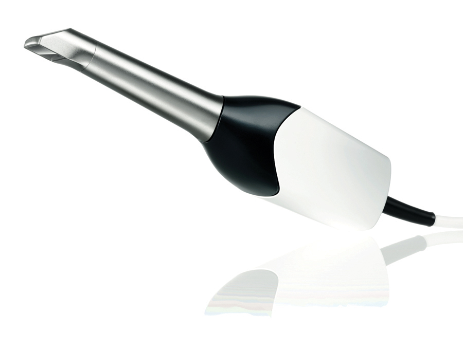
FIGURE 2. Colour Spectrum of the Omnicam

The Omnicam records whatever it “sees” and can image both the Maxillary and Mandibular arch (Fig. 3). In order to relate the arches together, a “buccal bite” image would be taken which is analogous to a bite registration. A buccal bite is created by having the patient close their mouth in full Centric Occlusion. An image of the teeth in Centric Occlusion is taken (Fig. 4) and utilized to create a virtual articulator. In creation of the virtual articulator, the models are placed virtually to define a model axis and to create a highly accurate proposal of the restoration.
FIGURE 3. Full Arch Imaging
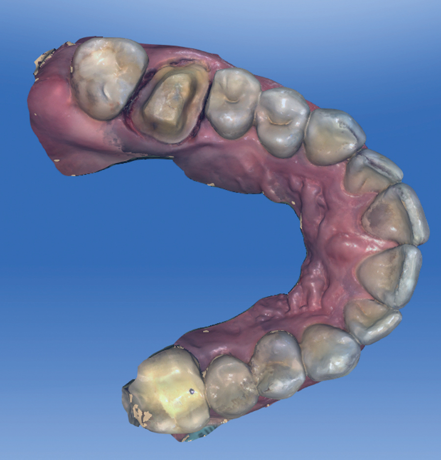
FIGURE 4. Buccal Bite Registration
Prior to virtually fabricating the restoration, the virtually mounted models must be assessed critically. At this point, we are critical of our own preparation for we can visually (quantitatively if desired) determine if we have a sufficient amount of reduction, a smooth surface finish, rounded internal edges and if are able to clearly visualize the margin on the virtual model. Fortunately, due to the fact that imaging is rapid to facilitate inspection, if there is insufficient reduction or a poor margin, then one can immediately go back to the oral cavity and correct the deficiency. Upon correction of the deficiency, one only needs to image that portion of the intra-oral cavity after first having “cut” that portion out of the model. The next step in the workflow is to define the margin of the restoration. By successfully retracting the margin prior to imaging, the margin of the restoration is easily defined (Fig. 5). After margination, the CEREC software will propose a restoration (Fig. 6). The proposal is accomplished with BioJaw. BioJaw is a tooth design algorithm that analyzes all of the adjacent teeth that are imaged to determine a mathematical morphology of the patient’s teeth. This individual fingerprint is then utilized to create an occlusal pattern that closely mimics the patient’s natural teeth. Alternatively, one can utilize the software to alter the tooth morphology to their choice.
FIGURE 5. Margination of the Preparation

FIGURE 6. Initial Proposal of the Restoration with a Colour Map of the Occlusal Contact Points on the Full Arch
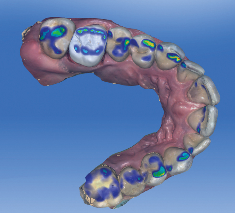
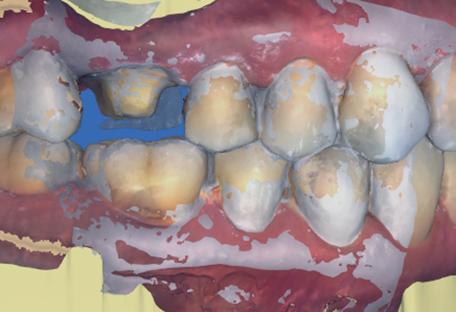
The virtual proposal of the restoration must be analyzed to ensure we have sufficient contact points with the adjacent teeth and an acceptable occlusal relationship with the opposing arch (Figs. 7 & 8). Regardless of the fact that the CEREC software will propose a restoration, which may require no adjustment by the user, the virtual proposal must be assessed critically. The restoration is analyzed by assessing the colour map on the restoration. This colour map indicates the proximity to the adjacent teeth, the opposing arch and the thickness that the restoration will be to ensure clinical success. The virtual restoration can be easily adjusted by utilizing design elements in the CEREC software.
FIGURE 7. Colour Map of the Occlusal Contact Points
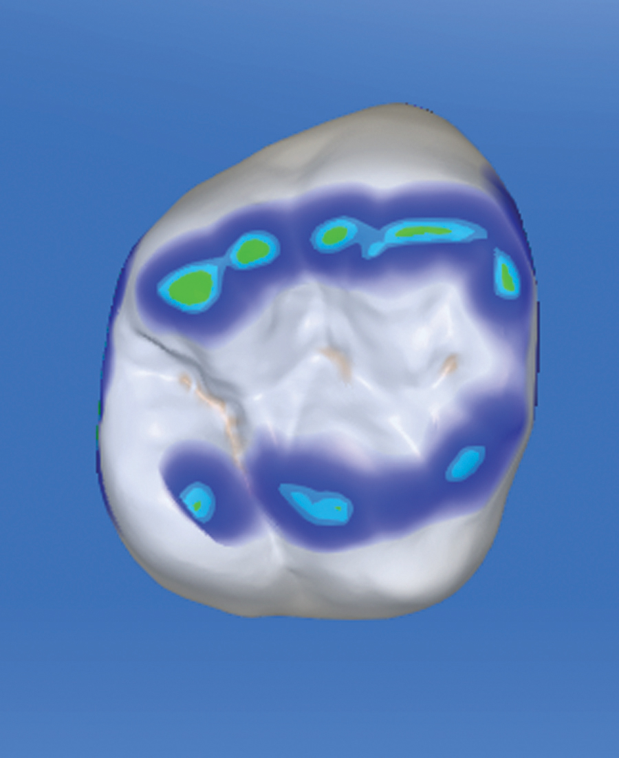
FIGURE 8. Colour Map of the Proximal Contact

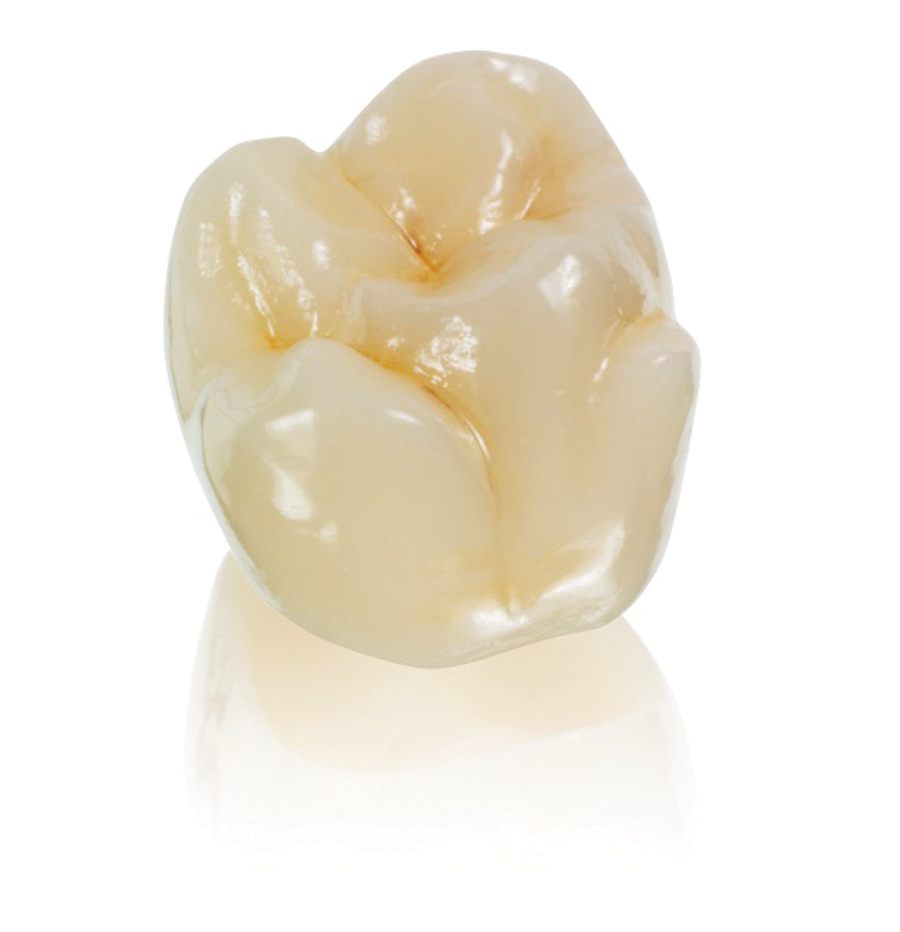
The fabrication of the restoration is accomplished rapidly in the office with a Milling Chamber, which occupies a very small footprint (Fig. 9). In fabrication of the restoration on the CAM system, a decision can be made on what type of dental material will be utilized. Fortunately, almost every restorative indication that can be ordered from a commercial laboratory can be selected at this stage. Dental materials such as e.MAX (lithium disilicate), CELTRA Duo (zirconia reinforced lithium silicate), Empress or Zirconia can be utilized, to name a few. These materials are available to us in a block format (Fig. 10), which is then milled in the Milling Chamber to create the restoration, which has been stained and glazed (Fig. 11). The choice of the restorative material is left to the discretion of the user. All of the materials can be fabricated chairside in one visit except for Zirconia. Due to the fact that Zirconia, as a material, needs to be Sintered in a Sintering Oven, it is best to have the patient return for insertion of that restoration. To ensure high flexural strength of the material and ensure great aesthetics, one can quickly glaze the material in a Porcelain Furnace. This utilization of the Porcelain Furnace serves two purposes. The first purpose is “healing” of the porcelain. During fabrication of the restoration and during the try-in of the restoration, small micro fractures may be introduced to the porcelain. The Porcelain Furnace raises the temperature of the dental material sufficiently that the micro-fractures coalesce into one homogenous structure and the restoration is healed. For a material such as e.MAX, the material needs to be crystallized so as to achieve optimal flexural strength. Use of the Porcelain Furnace can elevate the flexural strength of up to approximately 400 MPa.
FIGURE 9. Milling Chamber

FIGURE 10. Blocks of Different Restorative Materials

FIGURE 11. Final Restoration for Insertion
If the restoration contains a glass ceramic, hydrofluoric acid must be first utilized to create a micro-mechanical etching pattern. The surface of the restoration will then be silanated. Depending on the resin cement being utilized, the surface of the tooth will be prepared for bonding and the restoration inserted.
Digital Dentistry utilizing CEREC is an efficient method to create beautiful and strong restorations in one visit. The creation of these restorations in one visit saves time for both the practitioner and patient, as the patient does not have to return for a subsequent insertion visit.OH
Dr. Bobby Chagger is a graduate of the University of Toronto and has a strong interest in dental implantology utilizing ConeBeam and CEREC. He has successfully integrated CEREC and ConeBeam into his five ChaggerDental Offices in Southern Ontario, which are situated in the Cities of Mississauga, Oakville, Burlington and Dundas. His passion and insight have brought him international recognition as a Speaker and Educator. Look to claritywerx.com for more on digital dentistry.
Oral Health welcomes this original article.


