Introduction
Kissing teeth are extremely rare incidental radiological findings. Only very few cases have been reported in the literature. Kissing mandibular teeth can be divided into kissing molars (KM) and kissing canines (KC).
Kissing Molars
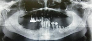
KM are impacted permanent molars that have occlusal surfaces contacting each other in a single follicular space with roots pointing in opposite directions (Boffano & Gallesio 2009) (Figure 1).
Figure 1

Panoramic scan showing third and fourth molars. In the lower right jaw are characteristic unilateral kissing molars (KM, class III).
“Kissing molars” were first descripted by Van Hoof in 1973. He reported a case of four impacted molars (two bilateral KM) in a 31-year-old patient (Van Hoof 1973). This special term was used again from Robinson et al. in 1991 (Robinson et al. 1991). In the present literature KM are also named as “rosette formation“ and “rosetting of molars“ (Menditti et al. 2015), or entitled as ”impacted love“ (Gulses et al. 2012).
KM often are retained, distally angulated second molar and a mesially angulated, displaced wisdom teeth. KM are an unusual form of impaction that is very infrequently reported in dental literature (Brauer 2017). KM can occur unilateral or bilateral (Robinson et al. 1991). Gulses et al. further divide in class I (first and second lower molar), class II (second and third lower molar), and the extremely rare class III (third and fourth supernumerary lower molar) (Gulses et al. 2012).
KM usually are asymptomatic and often an incidental finding in panoramic radiographs (Brauer 2017). The etiology and pathogenesis are unknown. However, the association to metabolic connective diseases such as mucopolysaccharidosis has been suggested (Menditti et al. 2015). Indications for surgery involve a history of pain, recurring infections, or cystic lesions (Arjona-Amo et al. 2016, Scheuber et al. 2014). The surgical approach for this condition requires a meticulous understanding of the anatomy of the region, advanced surgical abilities, and an individual rigorous planning process (Arjona-Amo et al. 2016, Boffano & Gallesio 2009).
Kissing Canines
Bilateral impacted mandibular canines when transmigrated can present as kissing phenomenon in the panoramic radiograph (Figure 2). Qaradaghi termed bilateral mandibular canines as KC or alternative as “mirror image canines” in cases which are defined as the migration of both canines at the same rate and on the same horizontal axis parallel to each other and meeting each other at the midline (Qaradaghi 2010).
Figure 2
KC can be associated with cystic formation and/or transmigration. Transmigration is usually defined as a phenomenon in which more than half of the length of an unerupted tooth has passed the midline.
There are very few cases of KC reported in the literature. For example, Sanjay et al. reported an unusual case of a 24-year-old female with kissing dentigerous cysts involving mandibular canines (Sanjay et al. 2015). Similarly, Dongol et al. described a case of bilateral mandibular canine impaction with transmigration in a 32-year-old male patient (Dongol et al. 2017) and Sharma et al. reported a case of KC with their labial surfaces facing each other and sharing a common follicular space in a 21-year-old man (Sharma et al. 2014). In contrast, Omami & Mathew reported a case of KC with a dentigerous cyst in a 12-year-old girl (Omami & Mathew 2015).
The etiology and pathogenesis are unknown. Management of KC includes periodic observation, surgical exposure and orthodontic alignment, transplantation, and surgical removal (Dongol et al. 2017, Sharma et al. 2014).
References:
- Arjona-Amo M, Torres-Carranza E, Batista-Crusado A, Serrera-Figallo MA, Crespo-Torres S, Belmonte-Caro R, Albisu-Andrade C, Torres-Lagares D, Gutiérrez-Pérez JL. Kissing molars extraction: Case series and review of the literature. J Clin Exp Dent 2016; 8(1): e97-e101
- Boffano P, Gallesio C. Kissing molars. J Craniofac Surg 2009; 20(4): 1269-1270
- Brauer HU. Kissing molars – Ein unerwarteter, seltener radiologischer Befund. ZWR 2017; 126(5): 245
- Dongol A, Acharya P, Yadav RP, Mahat AK, Yadav AK, Shrestha A, Jaisani MR. Kissing canines associated with dentigerous cyst, a case report of transmigrated bilateral mandibular canines. Int J Oral Craniofac Sci 2017; 3(1): 14-16
- A, Varol A, Sencimen M et al. A study of impacted love: kissing molars. Oral Health Dent Manag 2012; 11(4): 185-188
- Menditti D, Laino L, Cicciu M et al. Kissing molars: report of three cases and new prospective on aetiopathogenetic theories. Int J Clin Exp Pathol 2015; 8(12): 15708-15718
- Omami G, Mathew R. Kissing canines associated with a dentigerous cyst: case report and mini review. Int J Dentistry Oral Sci 2015; 2(6): 84-86
- Qaradaghi IF. Bilateral transmigration of impacted mandibular canines: report of two cases and review. Rev Clín Pesq Odontol 2010; 6(3): 271-275
- Robinson JA, Gaffney W Jr, Soni NN. Bilateral “kissing” molars. Oral Surg Oral Med Oral Pathol 1991; 72(6): 760
- Sanjay CJ, David CM, Kaul R, Shilpa PS. Kissing dentigerous cysts involving mandibular canines: report of unusual case with review of the literature. J Calif Dent Assoc 2015; 43(1): 29-33
- Scheuber S, Bornstein M. Kissing molars. A peculiar radiologic finding. Swiss Dent J 2014; 124(1): 16-17
- Sharma S, Raghavan V, Kumari S. Kissing mandibular canines: serendipity at its best. J Indian Acad Oral Med Radiol 2014; 226(1): 82-84
- Van Hoof RF. Four kissing molars. Oral Surg Oral Med Oral Pathol 1973; 35(2): 284.
Dr. Hans Ulrich Brauer is a Oral Surgeon, and Chief of Dentistry, Dorow Clinic Loerrach, Germany. He is also an editorial board member of the German dental journal ZWR.
Dr. Bruce Pynn is Oral Health’s editorial board member for oral and maxillofacial surgery. He is an Associate Professor, North Ontario School of Medicine, Lakehead University, and Chief of Dentistry, Thunder Bay Regional Health Science Center.