
 The GentleWave® Procedure effectively cleans and disinfects while conserving more precious tooth structure. Simultaneous cleaning of all canals, deep into dentinal tubules, isthmi, lateral canals and other complex anatomy is accomplished through a proprietary technology that leverages multisonic energy, advanced fluid dynamics, and accelerated chemistry for improved patient outcomes.
The GentleWave® System’s mechanism of action involves proprietary physicochemical phenomena which have been diligently invented and tuned to overcome the limitations posed by both the root canal system and standard root canal instrumentation. It therefore enables fast and thorough cleaning and disinfection of a highly complex and unpredictable root canal space encapsulated with porous dentin which can harbor millions of bacteria.
The degassed procedure fluid is both the initiator and conduit of the entire procedure. This fluid is used to generate a broad spectrum of acoustic energy, is the medium delivering the acoustic energy throughout the root canal and is also deflected to induce gentle vortices and slight negative pressure inside the root canals. Additionally, vapor lock, one of the main challenges in conventional irrigation which can significantly hinder cleaning and disinfection, is addressed by simply reducing the gas content of the procedure fluids and a simple sealed platform. A broad spectrum of acoustic energy ensures efficient delivery to all anatomical features of the root canal system, with various length scales ranging from tubules to anastomoses to main canals.
Sonendo’s tooth-saving technology has been designed and developed specifically for endodontics, as an alternative to standard root canal treatment, and with a mission of helping you save more teeth for more patients through Sound Science.®
A Few Key Features that Set the Gentlewave® Procedure Apart
- The procedure fluids are optimized in the GentleWave® Console through a process known as degassing. This reduces the possibility of apical vapor lock and maximizes tissue dissolution, debridement, and disinfection.
- With the GentleWave® Procedure, the job of the file is simply to provide an uninterrupted path for procedure fluids and a 3-dimensional fill.
- A sealed environment is generated between the tooth and the GentleWave® Procedure Instrument to allow for constant refreshing of degassed procedure fluids and simultaneous evacuation of debris.
- Operating within an air-free, sealed root canal system, filled only with procedure fluids, is key.
- It’s in the Procedure Instrument that patented Multisonic Ultracleaning® technology enters the equation. The stream of optimized fluid entering the tooth creates a powerful shear force, which causes hydrodynamic cavitation in the form of a useful cavitation cloud. The implosion of thousands of micro-bubbles creates an acoustic field of broadband frequencies which travels through the fluid into the entire root canal system.
- Literature states that the pressure in the periapical region is positive.1,2 With the GentleWave® System, gentle vortical flow is induced within the root canal system with a slight negative pressure at the apices which reduces the potential for extrusion. Together, the GentleWave® System and periapical region create a pressure differential. Maintaining this pressure differential is essential for the GentleWave® System Multisonic Ultracleaning technology.
The result, unique to the GentleWave® Procedure, is unmatched cleaning and disinfection from the crown to the apex.
Six-Month Healing of a Mandibular First Molar with Complex Anatomy
Courtesy of Khang T. Le, DDS (Figs. 1-6)
History
A 30-year-old male was referred for evaluation to a private endodontic clinic. The patient had a chief complaint of spontaneous pain on the left side of the mouth.
Diagnosis
Radiographic examination of the mandibular left first molar (#19) revealed a large filling on the occlusal and distal aspects of the tooth. Radiolucency was seen around both roots of the subject tooth with a larger lesion on the mesial root. Clinical examination revealed a fracture line on the distobuccal cusp. The subject’s tooth exhibited mild pain response to both palpation and percussion as well as class I mobility. The response of the tooth to cold thermal pulp vitality test was negative. After clinical and radiographic examinations, the tooth was diagnosed as necrotic pulp with symptomatic apical periodontitis.
Gentlewave® Procedure
The patient was anesthetized and the tooth was isolated with a rubber dam. Caries and existing restoration were removed. Conservative straight-line access was performed and clinical examination revealed three distinct orifices: two located mesially and one distally. Instrumentation was accomplished by shaping the canals up to size #15 K-file and using only two NiTi rotary files for a final apical size of #20. Irrigation with sterile water was used between each instrument to prevent the build-up of debris. Orifice openers were not utilized to conserve dentinal structure within the coronal third. The GentleWave® Procedure was utilized after instrumentation to remove pulp tissue remnants, debris, smear layer and bacteria.
All root canals were obturated using a warm vertical condensation technique with gutta-percha and AH Plus® sealer. After obturation, a complex root canal system was visible with two inter-canal isthmi and a lateral canal. The patient reported no discomfort during or after the GentleWave® Procedure. The post-GentleWave® Procedure radiograph shows the mandibular first molar complex root canal system with at least two visible inter-canal isthmi and a lateral canal.
At the three-month recall, the radiograph shows evidence of healing with a reduction in the size of the periradicular lesion. At the six-month recall, radiographs revealed complete healing of apical periodontitis. The time-lapse radiographs of healing up to 18 months after the GentleWave® Procedure provide verification of healing.
Fig. 1
Pre-procedure.

Fig. 2
Post-procedure.

Fig. 3
Three-months post-procedure.
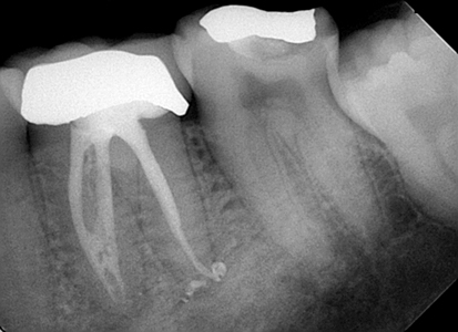
Fig. 4
Six-months post-procedure.

Fig. 5
12-months post-procedure.

Fig. 6
18-months post-procedure.

Healing After Silver Point Retreatment with the GentleWave® Procedure
Courtesy of Randy W. Garland, DDS (Figs. 7-12)
History
A 71-year-old female presented with biting tenderness in the lower right quadrant. The patient’s medical history was noncontributory.
Diagnosis
Radiographic examination revealed prior silver point root canal therapy with periapical lesions on the mesial and distal roots of the lower right first molar (#30). Clinical examination showed tenderness to bite percussion and palpation. Based on the clinical and radiographic findings, a diagnosis of acute apical periodontitis was made.
Gentlewave® Procedure
The patient was anesthetized and the tooth was isolated with a rubber dam. A conservative straight-line access was prepared and the silver points were removed utilizing ultrasonics and Hedstrom files. The GentleWave® Procedure was determined to be the best treatment modality to prevent reinfection and remove bacteria after silver point removal. Instrumentation was completed with only two NiTi files to provide a fluid and obturation path prior to the GentleWave® Procedure.
After the GentleWave® Procedure, the paper points utilized to dry each canal were completely clean when removed. There was no presence of black sediment as typically seen post-standard silver point root canal retreatment.
Obturation was completed using a warm vertical technique with gutta-percha and AH Plus® sealer.
The patient reported no discomfort during or after the procedure.
At the nine-month follow-up, clinical evaluation showed the tooth was asymptomatic. Radiographic evaluation showed significant bone regrowth and periradicular healing.
Fig. 7
Pre-procedure radiograph.

Fig. 8
Pre-procedure CBCT.

Fig. 9
Post-procedure.

Fig. 10
Post-procedure CBCT.

Fig. 11
Silver points removed.

Fig. 12
Nine-months post-procedure.

Nine-Month Healing of a Mandibular Molar with an Uninstrumented Middle Mesial Canal
Courtesy of Stacey M. Woo, DDS, PhD (Figs. 13-16)
History
A 25-year-old male with a non-contributory medical history presented to the clinic with a chief complaint of occasional mild sharp pain when biting and chewing.
Diagnosis
Clinical examination of the left mandibular molar (#19) revealed gross caries extending into the pulp and no Sensitivity to percussion or palpation. There was no response to vitality testing with Endo-Ice®. Pre-operative radiographic analysis showed periapical lesions on both mesial and distal roots. Based on clinical and radiographic findings, the diagnosis was pulpal necrosis and asymptomatic apical periodontitis.
Gentlewave® Procedure
The patient was anesthetized and the tooth was isolated with a rubber dam. A glide path was created with K-files up to size #20. Canals were instrumented with two NiTi rotary files for a final apical diameter of 20. Throughout instrumentation, 0.5% sodium hypochlorite and lubrication were used. The GentleWave® Procedure was utilized after instrumentation to remove pulp tissue remnants, debris, smear layer and bacteria.
All root canals were obturated using a warm vertical condensation technique with gutta-percha and AH Plus® sealer. Post-obturation radiographs show complex anatomies in the root canal system that were not previously visualized or instrumented, including a middle mesial canal and isthmi in the mesial and distal canals filled with obturation materials.
At the six-month recall, the patient was asymptomatic, and the tooth had been restored with a full coverage crown. The tooth was functional. Radiographic analysis showed healing of the periapical lesions. Complete resolution of apical periodontitis was noted at the nine-month recall and no clinical signs or symptoms were present.
Fig. 13
Pre-procedure.

Fig. 14
Post-procedure.

Fig. 15
Six-months post-procedure.

Fig. 16
Nine-months post-procedure.

2018
Peer Reviewed Publications
- Wang Z, Shen Y, Haapasalo M. Root Canal Wall Dentin Structure in Uninstrumented but Cleaned Human Premolars: A Scanning Electron Microscopic Study. J Endod. 2018 Mar (in press).
- Sigurdsson A, Garland RW, Le KT, Rassoulian SA. Healing of Periapical Lesions after Endodontic Treatment with the GentleWave Procedure: A Prospective Multicenter Clinical Study. J Endod. 2018;44(3):510-517.
- Ford MW. Utilizing the GentleWave® System for Debridement of Undetected Apical Anatomy. J Contemp Dent Pract. 2018;19(3):345-351.
Trade Journals
- Garland RW. Rapid Healing of a Large Periapical Premolar Lesion. Dentistry Today. 2018 [accepted].
2017
Peer Reviewed Publications
- Woo SM. Periapical Healing of a Mandibular Molar with Middle Mesial Canal: A Case Report. Journal of Interdisciplinary Medicine and Dental Science. 2017;5:209.
- DiVito EE, Le KT. Maxillary Molar Healing After Treatment of an Uninstrumented Canal with a Novel Root Canal Procedure: A Case Report. Clin Case Rep. 2017;5(10):1676-1681.
- DiVito EE, Rassoulian SA. Ex Vivo Scanning Electron Microscopy Evaluation of Cleaning Efficacy Following In Vivo Endodontic Treatment: A Report of 2 Cases. Dentistry. 2017;7:419.
- Ford MW. Complex Apical Anatomy Revealed Following Endodontic Treatment of a Maxillary Molar Using the GentleWave System: A Case Report. Dentistry. 2017;7:446.
- Garland RW. Bone Regrowth and Healing of Periapical Lesions Nine Months after Removal of Silver Points and Retreatment with the GentleWave Procedure. Int J Dent Oral Health. 2017;3(4).
- Pullen RV. Root Canal Treatment of a Maxillary First Molar with an Un-instrumented 5th Canal: A Clinical Case Report. J Oral Hyg Health. 2017;5:219.
Trade Journals
- Le KT. 18-month Case Study of a C-Shaped Mandibular Molar: Preserving Dentin and Deep Cleaning Utilizing an Innovative Procedure. Roots – The International C.E. Magazine of Endodontics. 1;2017.
- Advances in Root Canal Cleaning Help Improve Endodontic Outcomes. Decisions in Dentistry. April 2017.
- Keep it Real with Today’s Patients. Endodontic Practice US. Spring 2017.
2016
Peer Reviewed Publications
- Sigurdsson A, Garland RW, Le KT, and Woo SM. Twelve-Month Healing Rates after Endodontic Therapy Using the Novel GentleWave System: A Prospective Multicenter Clinical Study. J Endod. 2016; 42(7):1040-48.
- Sigurdsson A, Le KT, Woo SM, Rassoulian SA, McLachlan K, Abbassi F, Garland RW. Six-Month Healing Success Rates after Endodontic Treatment using the Novel GentleWave™ System: The PURE Prospective Multi-Center Clinical Study. J Clin Exp Dent. 2016;8(3):e290-8.
- Charara K, Friedman S, Sherman A, Kishen A, Malkhassian G, Khakpour M, Basrani B. Assessment of Apical Extrusion during Root Canal Irrigation with the Novel GentleWave System in a Simulated Apical Environment. J Endod. 2016;42(1):135-9.
- Le K. Six-month Healing of a Mandibular First Molar with Complex Anatomy Using a Novel Endodontic Procedure. J Dent Oral Disord Ther. 2016;4(4):1-3.
- Vandrangi P. Evaluating Penetration Depth of Treatment Fluids into Dentinal Tubules Using the GentleWave System. Dentistry. 2016; 6:366.
- Wang Z, Maezono H, Shen Y, Haapasalo M. Evaluation of Root Canal Dentin Erosion after Different Irrigation Methods Using Energy-dispersive X-ray Spectroscopy. J Endod. 2016;42(12):1834-1839.
- Haapasalo M, Shen Y, Wang Z, Park E, Curtis A, Patel P, Vandrangi P. Apical pressure created during irrigation with the GentleWave™ system compared to conventional syringe irrigation. Clinical Oral Investigation. 2016;20(7):1525-34.
Trade Journals
- Defining Our Terms. Refining Endodontics. Sonendo® Discusses the Mechanism Behind its New Standard of Clean. Endodontic Practice US. Winter 2016.
- The GentleWave System by Sonendo: Clean and Disinfect Even Complex Root Canal Anatomies with Sound Science – And a Revolutionary Mechanism of Action. Endodontic Practice US. Summer 2016.
- Is Your Practice Good Enough? Sonendo Discusses a System for Disinfection and Cleaning with Minimal Instrumentation. Endodontic Practice US. Spring 2016.
- Krupp J. Endodontic Advancements: How Leading Technologies Help Transform Endodontic Treatment Options. Dental Town. January 2016.
2015
Peer Reviewed Publications
- Ma J, Shen Y, Yang Y, Gao Y, Wan P, Gan Y, Patel P, Curtis A, Khakpour M, Haapasalo M. In Vitro Study of Calcium Hydroxide Removal from Mandibular Molar Root Canals. J Endod. 2015;41(4):553-8.
- Vandrangi P, Basrani B. Multisonic Ultracleaning in Molars with the GentleWave System. Oral Health. 2015;72-86.
- Molina B, Glickman G, Vandrangi P, Khakpour M. Evaluation of Root Canal Debridement of Human Molars Using the GentleWave System. J Endod;41(10):1701-5.
- Wohlgemuth P, Cuocolo D, Vandrangi P, Sigurdsson A. Effectiveness of the GentleWave System in Removing Separated Instruments. J Endod. 2015;41(11):1895-8.
Trade Journals
- Implementing the GentleWave System by Sonendo. Endodontic Practice US. January/February 2015.
- Ultracleaning of a Necrotic Molar Tooth After the Use of a Single Rotary File. Endodontic Practice US. March/April 2015.
- Sonendo: Challenging the Standard of Care with the GentleWave System. Roots – The International C.E. Magazine of Endodontics. 3;2015.
- Sonendo and Transformative Product Development: An Interview with Joseph Maggio, DDS. Endodontic Practice US. May/June 2015.
- Setting the Highest Possible Standard in Endodontics. Roots – The International C.E. Magazine of Endodontics. 2; 2015.
- Are you Transforming Endodontics? Endodontic Practice US. September/October 2015.
- New Horizons in Endodontics. Dental Advisor. November 2015.
- Resolve to Save More Teeth. Endodontic Practice US. November/December 2015.
Sonendo Case Reports
- Le KT. Signs of Healing Visualized within 6 Months: A Single- Visit Endodontic Treatment. Sonendo. 2015.
- Baker TF. Practicing Minimal Endodontics. Sonendo. 2015.
2014
Peer Reviewed Publications
- Haapasalo M, Wang Z, Shen Y, Curtis A, Patel, Khakpour M. Tissue Dissolution by a Novel Multisonic Ultracleaning System and Sodium Hypochlorite. J Endod. 2014;40(8):1178-81.
Trade Journals
- A New Paradigm in Endodontics. Endodontic Practice US. January/February 2014.
- Sonendo Aims to Transform the Future of Endodontics. Endodontic Practice US. March/April 2014.
- Sonendo: Root Canals Clean at the Speed of Sound. Roots – The International C.E. Magazine of Endodontics. 2; 2014.
- The Steps to Using the GentleWave System. Endodontic Practice US. November/December 2014. OH
Oral Health welcomes this original article.
References
- Cuicchi B, et al. Dental Fluid Dynamics in Human Teeth, In Vivo. J Endod. 1995;21:191.
- Stenvik A. Tissue Pressure and Histology of Normal and Inflamed Tooth Pulps in Macaque Monkeys. Arch Oral Biol. 1972;17:1501.
About the Author
 James A. Smith, Jr., D.M.D., received his dental degree in 1980 from the University of Alabama School of Dentistry and completed his endodontic residency there in 1982. He has been in private endodontic practice for almost 35 years and is an adjunct assistant professor with the Department of Endodontics at The University of Alabama at Birmingham School of Dentistry. Dr. Smith has co-authored an article that was published in the 2015 Journal of Endodontics. His practice is located in Birmingham, Alabama.


