
Technology is changing the way we live and work, improving our lives. Smart phones and watches keep us connected to the world, monitor our heart rates, detect and alert atrial fibrillation, check glucose levels, count our steps, record our hours of sleep and keep an eye on numerous vital statistics (Fig. 1). Devices respond and react to our voices, controlling our environment and our safety, giving guidance, arranging transportation and summoning emergency help.
Fig. 1

Technology has also transformed the modern dental office. Treatment is tracked electronically, patient data and health history are stored and accessed remotely. Communication and patient education are provided on mobile electronic devices. Patient telephone calls instantly display caller photos, family members, unscheduled treatment and special messages about their care (Weave, Ogden, UT) (Fig. 2). Digital photography, intra-oral impression scanners, 2-dimensional digital radiography and 3-dimensional CBCT scans are used to evaluate and communicate our patient’s oral condition (Fig. 3). These images are readily shared with other associated professionals (Fig. 4).
Fig. 2

Fig. 3

Fig. 4

Minimally invasive medical treatment is now the standard of care. Preservation of original tissue is paramount; diagnosing pathology at its earliest stages reduces the severity of treatment. Technological advances allow us to detect and confirm disease sooner.
Regular oral cancer screening is one of the most important services that dental practitioners provide for their patients. Dentists are the physicians of the mouth, generally seeing patients more frequently and systematically than any other healthcare provider.
Innovative screening devices improve our ability to detect suspicious lesions. They alert the dentist to concerns that may not be readily visible. Dysplastic and hyper-plastic tissue contains metabolic intermediaries that block normal tissue fluorescence.¹ The VELscope VX (LED Medical Diagnostics, Vancouver, BC) employs a 400-460nm light which fluoresces healthy tissue (Fig. 5). The BIO/SCREEN (Addent, Danbury, CT) uses 5 LED lights which emit 400-450nm light (Fig. 6). When observed through a dedicated scope, non-fluorescing (or potentially abnormal) areas appear dark (Fig. 7). While not offering a definitive diagnosis, these devices are excellent screening devices to alert the dentist to potential pre-cancerous or cancerous tissues.²
Fig. 5

Fig. 6

Fig. 7

Digital dental photography is invaluable in documenting questionable areas and sharing them with patients and specialists. The Shofu EyeSpecial C-III (Shofu, San Marcos, CA) is a simplified, lightweight, digital dental camera that allows all dental team members to easily take intra-oral photos (Fig. 8). All too often, patients do not see a specialist for a follow-up evaluation. Waiting six months or more for the next periodic dental appointment may prove fatal with some aggressive lesions. Taking digital photos and sending them to a pathologist with a written narrative is a quick and verifiable method for ensuring follow-up (Fig. 9). The specialist may decide that the area is benign or that it needs immediate attention. Our practice has been the first detector of suspicious areas that were referred to specialists and diagnosed as HIV on three occasions. Early treatment can significantly extend the lifetime of a patient.
Fig. 8
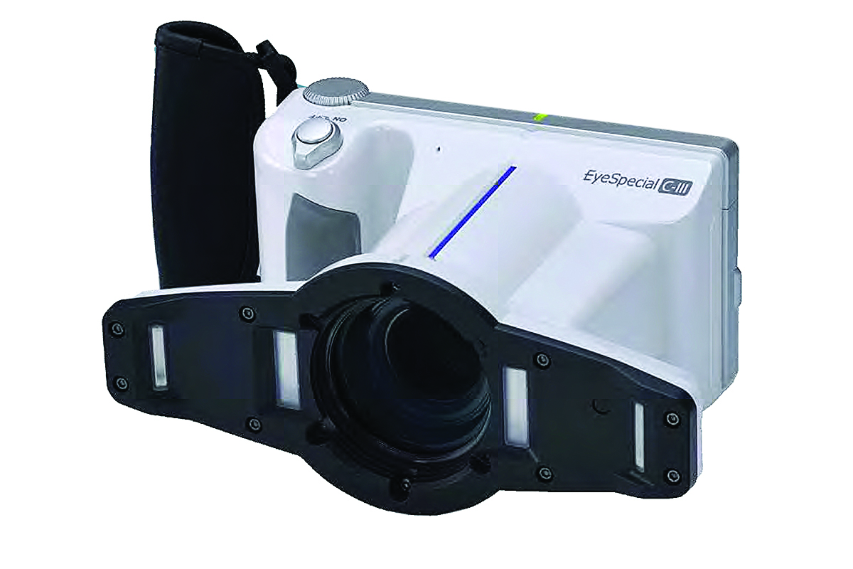
Fig. 9

Traditional caries detection methods (mirror, explorer and radiographs) are well-established, but sometimes inconclusive. Studies have shown that with conventional visual/tactile techniques, a significant number of occlusal carious lesions are overlooked.³ Significant cumulative destruction may occur before detection by conventional methods. Laser devices can add valuable diagnostic information. Carious lesions, when exposed to certain wavelengths of light, emit more intense fluorescence than sound tissue due to organic components and protein chromophores found in affected tooth structure.4 Fluorescence induced by 655 nm red light has been shown to effectively differentiate between sound and carious tooth structure. The Diagnodent (Kavo/Kerr, Orange, CA) emits red laser light and then measures the intensity of the reflected fluorescence, providing a numeric reading (Fig. 10). The higher the number, the more extensive the decay. The device emits a sound that increases in intensity as the numbers increase, reinforcing to the patient the potential severity of the lesion. While unable to definitively diagnose caries extent, the unit is an excellent adjunct in diagnosis and treatment planning.5
Fig. 10

Fluorescent Class I Case Presentation
A suspicious occlusal pit was visually noted on a young patient in for their periodic examination (Fig. 11). The explorer examination was inconclusive. The area was cleaned and the Diagnodent calibrated and placed over the questionable area (Fig. 12). The reading of 42 indicated the likelihood of caries. An erbium based hard tissue laser (Lite Touch, Yokneam, Israel) (Fig. 13) was used to excavate the area without anesthesia, very advantageous for young, apprehensive patients. The entire preparation was performed by the laser (Fig. 14). Small excavations make thorough void-less fillings difficult. High viscosity (heavily filled) flowable composites are strong enough to withstand occlusal forces, easy to apply into small preparations, and have excellent translucency and polishability. Beautifill Flo XR (Shofu, San Marcos, CA) is a high viscosity flowable composite Giomer with pre-set glass-ionomer filler particles that offer true fluoride release (Fig. 15). The composite resin material was placed, shaped, finished and polished (Fig. 16).
Fig. 11

Fig. 12

Fig. 13

Fig. 14

Fig. 15

Fig. 16

Computerized trans-illumination devices assist with difficult-to-detect interproximal lesions. While digital radiographs can be enlarged, lightened/darkened and the contrast adjusted, interproximal lesions may still be difficult to diagnose. The Cari-Vu (Kavo/Kerr, Orange, CA) is an advanced intra-oral trans-illumination monitor (Fig. 17). The handpiece is placed around the tooth; one side emits a specific wavelength that enhances caries detection, while the other side houses a camera connected to a monitor. Decay can be viewed on the screen and stored in the computer. Though the device does not eliminate the need for radiographs, it is an excellent adjunctive method to help reach a more definitive diagnosis.6

Trans-illumination Class II Case Presentation
Four bitewing radiographs were taken as part of a periodic oral examination. Evaluation revealed a possible interproximal carious lesion on the maxillary left first molar (Fig. 18). Visual examination did not clarify the diagnosis (Fig. 19). The Cari-Vu handpiece was positioned to clearly show the area in question. The image was captured and appeared on screen, clearly delineating the carious area and its extent (much larger than anticipated from the radiographs and visual examinations) (Fig. 20). Operative treatment was scheduled.
Fig. 18

Fig. 19

Fig. 20

The patient was anesthetized, and the occlusal approach allowed access to the decay (Fig. 21). A slow-speed round bur removed the remaining diseased tissue. Fuji-Liner (GC, Alsip, IL), a resin-modified glass ionomer was placed in the deepest areas (Fig. 22). After the sectional matrix, Optibond eXTRa (Kavo/Kerr, Orange, CA) universal dentin bonding agent was applied (Fig. 23). This two-component system effectively etches enamel without phosphoric acid; component one is a hydrophilic primer, and component two is a hydrophobic adhesive. Together, they create a stable bond less likely to suffer from hydrolytic degradation.7 Then, Sonic-Fill III (Kerr/Kavo, Orange, CA), a unique bulk-fill composite that significantly reduces polymerization shrinkage stress was introduced (Fig. 24). Sonic energy is applied to the composite as it is dispensed, initially deceasing its viscosity and delaying the gel state upon light exposure. This reduces polymerization shrinkage stresses that can be detrimental to tooth structures.8 The cavity can be filled in a single step as the bulk-fill has expanded curing depth and reduced shrinkage stress. Sonic-Fill III facilitates posterior composite placement; the tip is placed into the box and the foot pedal depressed. The composite was placed in one single increment and polymerized occlusally for 20 seconds with the Demi Plus (Kavo/Kerr Orange, CA) high output curing light. The bulk-fill technique requires adequate curing of the entire composite restoration, particularly in the deep interproximal box. A high-output curing light with a collimation pattern that ensures adequate deep curing used for the proper length of time is essential9 (Fig. 25). The sectional matrix ring was removed and the wings of the matrix peeled back to allow light curing at the gingival areas from both buccal and lingual. Finally, the matrix was removed, and the tooth polymerized 20 seconds each from the occlusal, buccal, and lingual. The restoration was shaped, finished and polished. The tooth was post-cured for an additional 20 seconds (Fig. 26). The ability to confidently and predictably place large direct Class II composite restorations has dramatically increased with the use of sectional matrices, universal bonding agents, bulk-fill composites and high-output LED curing lights.

Fig. 22

Fig. 23

Fig. 24

Fig. 25

Fig. 26

Conclusion
New dental technology has improved care for our patients. New devices help detect pathology sooner allowing less invasive treatment. New restorative systems improve their durability and simplify the placement of direct composites, making it easier for dentists to use more conservative preparations rather than full-reduction crowns. The adage that “Dentistry begets Dentistry” has never been more relevant. With patients living longer, we need to use minimally invasive techniques to allow for the retention of maximum tooth structure to ensure that teeth will last a long lifetime.
Oral Health welcomes this original article.
References
- Mascitti M, Orsini G, Tosco V, Monterubbianesi R., et al. An Overview on Current Non-invasive Diagnostic Devices in Oral Oncology. Front Physiol. 2018;25(9):1510.
- Poh C. F., Zhang L., Anderson D. W., Durham J. S., Williams P. M., Priddy R. W., et al. Fluorescence visualization detection of field alterations in tumor margins of oral cancer patients Clin. Cancer Res. 2006;12:6716–6722.
- Braga MM, Mendes FM, Ekstrand KR. Detection activity assessment and diagnosis of dental caries lesions. Dent Clin North Am. 2010;54(3):479-493.
- Carvalho F, Barbosa, A, Zanin F, Fátima A, et al. Use of laser fluorescence in dental caries diagnosis: a fluorescence x biomolecular vibrational spectroscopic comparative study. Braz. Dent. J. 2013;24:59-63.
- Khalife MA, Boynton JR, Dennison JB, Yaman P, Hamilton JC. In Vivo Evaluation of DIAGNOdent for the Quantification of Occlusal Dental Caries. Operative Dentistry. 2009, 34-2, 136-141.
- Litzenburger F, Heck K, Pitchika V, Neuhaus KW, Jost FN, Hickel R, Jablonski-Momeni A, Welk A, Lederer A, Kühnisch J. Inter- and intraexaminer reliability of bitewing radiography and near-infrared light transillumination for proximal caries detection and assessment. Dentomaxillofac Radiol. 2018;47(3):20170292.
- Tjäderhane L, Nascimento, F, Breschi L,Mazzoni. A Optimizing dentin bond durability: strategies to prevent hydrolytic degradation of the hybrid layerDent Mater. 2013 Oct; 29(10): 999–1011.
- Chesterman J, Jowett A, Gallacher A, Nixon P. Bulk-fill resin-based composite restorative materials: a review. Br Dent J. 2017;222:337-344.
- Price R, Shortall A, Palin W. Contemporary Issues in Light Curing. Oper Dent.2014; 39(1): 4-14.
About the Author
 Dr. Ward is in private practice in Columbus, Ohio. He is a Diplomat of the American Board of Aesthetic Dentistry, a Fellow of the American Society for Dental Aesthetics, Fellow in the Academy of General Dentistry, Fellow in the International Academy for Dental-Facial Esthetics and Fellow in the American and International College of Dentists. He has lectured internationally and authored numerous articles in the field of proportional smile design. Dr. Ward can be contacted at dward@columbus.rr.com.
RELATED ARTICLE: 3D Technology In The Dental Practice
Follow the Oral Health Group on Facebook, Instagram, Twitter and LinkedIn for the latest updates on news, clinical articles, practice management and more!


