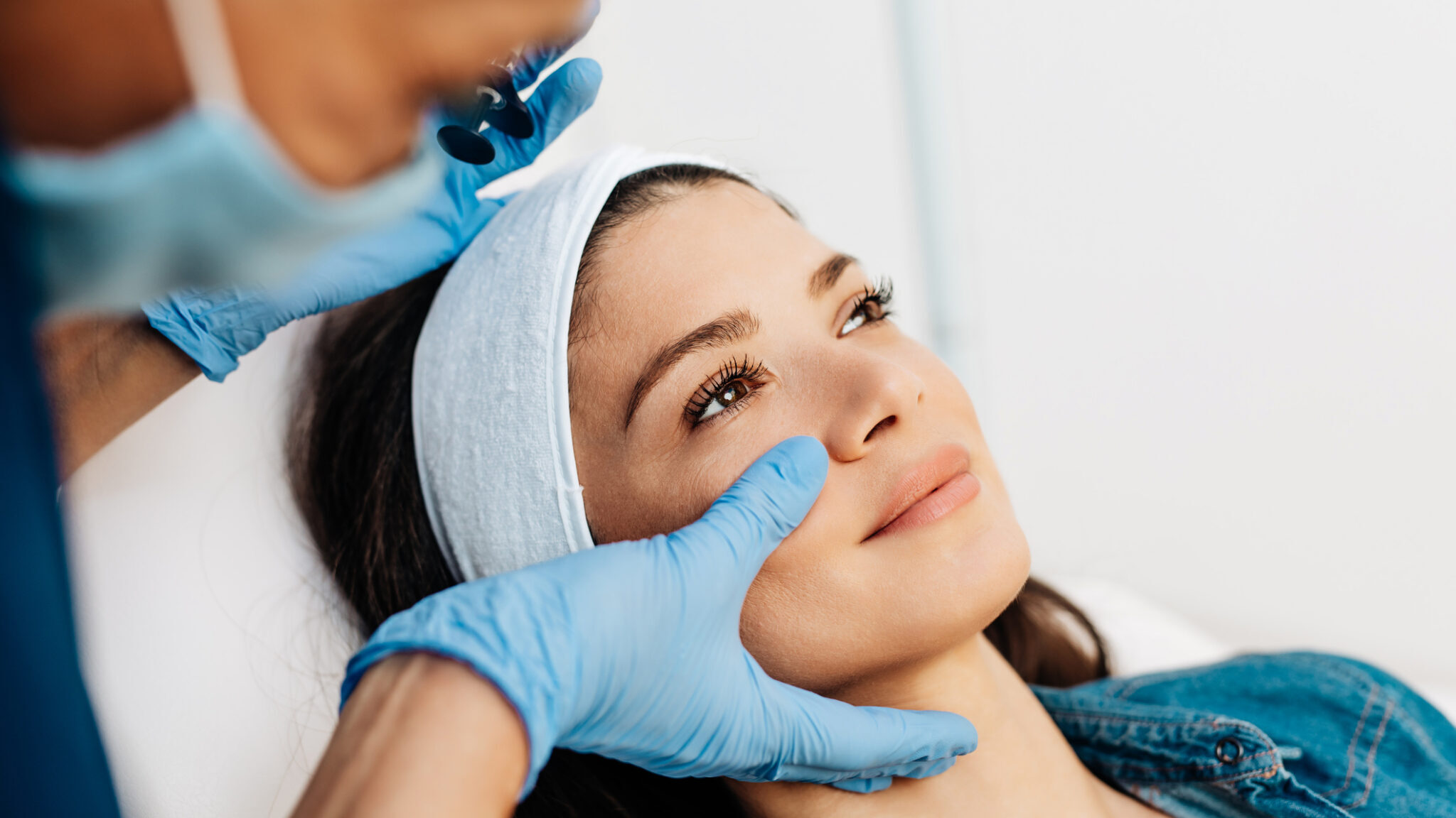
What does the word “Botox” conjure up in your mind? A “frozen”, unnatural facial expression? Unnecessary treatment that caters to the vain and is provided by those seeking to line their own pockets? A “poison” that is dangerous and should not be used, especially by dentists? If these descriptives pop into your mind then a paradigm shift is in order. The art and science of injecting Botulinum Toxin Type A (aka “Botox”, Dysport”, “Xeomin” etc.) is a safe and effective treatment for numerous conditions that a properly trained dentist is ideally suited to administer.
Historical Background
Botulinum Toxin Type A (scientifically known as BoNT-A) entered the cosmetic world in 1991 when Drs. Jean and Alastair Carruthers presented their findings at The American Society for Dermatologic Surgery in Orlando, Florida. It was met at the time by an underwhelming response from their colleagues. Dr. Jean Carruthers, a Vancouver-based ophthalmologist, had been one of the first in Canada to use Botox for medical purposes starting in 1983. While using it to treat blepharospasm, a condition that causes uncontrollable blinking of the eye, she noticed that the injections resulted in reduction of peri-orbital rhytids, so-called “crow’s-feet”. Along with her husband Dr. Alastair Carruthers a dermatologist, they began injecting forehead worry or frown lines, linking the therapeutic use of the drug with a cosmetic use.
Mechanisms of Action
The primary mechanism of action had earlier been identified as muscle relaxation due to the prevention of the release of the neurotransmitter acetylcholine from the efferent nerve and its associated muscle. In other words, BoNT-A prevented the nerve from telling the muscle to contract, relieving the blepharospasm. Orbicularis oculi encircles the eye and its lateral fibres were serendipitously also affected by the drug when injected to relieve blepharospasm, rendering it unable to strongly contract and produce “crow’s-feet”. A therapeutic treatment had inadvertently resulted in a positive cosmetic side effect. Soon other muscles were being targeted for relaxation and today there are over eight hundred medical uses for BoNT-A including treatment of cervical dystonia, hyperhidrosis, overactive bladder, amblyopia or “lazy eye”, and chronic migraines to name a just few (see Time Magazine 2017-01-18, Fig. 1).
Fig. 1

As dentists we are confronted daily with patients experiencing varying degrees of pain and dysfunction. Head and neck pain, temporomandibular joint pain and dysfunction, facial muscle soreness along with tooth wear and loss are common complaints. Many modalities are used to treat the causes or relieve the symptoms: drugs, splints and orthotics, orthodontic treatment, occlusal adjustment, occlusal/restorative rehabilitation etc. BoNT-A is another safe and effective tool that can be added to the dentist’s tool box.
There are three mechanisms of action of BoNT-A that are responsible for relieving the headaches that often accompany temporomandibular disorders, headaches and migraine. The first, mentioned earlier, is the prevention of the release of the vesicle containing the messenger molecule acetylcholine at the neuromuscular junction, preventing the contraction of the associated muscle. Relaxation of hyperactive muscles is a major contributor to the pain relief that follows BoNT-A injections. However, it is not the only factor. There are two other mechanisms of action that contribute also.
The secondary mechanism involves prevention of the release of noxious neuropeptides such as “Substance P” involved in pain perception, vasodilation and neurogenic inflammation. “Substance P” is released from sensory nerve fibres in response to noxious stimuli and helps transmit pain afferent signals to the spinal cord and brain where the pain is perceived. BoNT-A inhibits its release.
The tertiary mechanism of action is directly on the central nervous system (CNS) where it affects the release of several neurotransmitters from central neurons. Passage of the afferent message to the CNS is accomplished via nerves transversing the cranial sutures. Various sutures lie beneath the sites in the temporalis and frontalis muscles that are often injected to treat headaches and migraine. The tertiary mechanism also works indirectly on the CNS by altering peripheral and central desensitization.
Cosmetic applications of BoNT-A generally involve the primary mechanism of action, namely muscle relaxation. However therapeutic applications involving pain also draw on the other two mechanisms.
Cosmetic Uses of BoNT-A
Upper Face
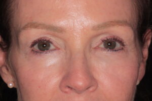
Treatment of the upper face provides the biggest cosmetic benefit for many people. Relaxing vertical frown or worry lines between the brows, horizontal forehead lines and crow’s-feet around the eyes are where the majority of treatment occurs (Figs. 2-10). In a younger person, these lines can often be prevented or eliminated. In a more mature person whose lines have become “static” (present even when the muscle is not contracting) the lines will be softened rather than eliminated. Including a brow lift to raise the corner of the eyebrow and make the eye appear more open (or a modified brow lift that also targets some of the headache-causing temporalis muscle) is an additional means of increasing cosmetic benefit. Cosmetic treatment of upper face areas (apart from crow’s-feet) often has a significant impact on relieving headaches, providing a prime example of the synergy between cosmetic and therapeutic treatment.
Fig. 2

Fig. 3

Fig. 4

Fig. 5

Fig. 6

Fig. 7

Fig. 8

Fig. 9

Fig. 10

Cosmetic uses for BoNT-A within the scope of dentistry include improving the frame around a smile makeover (the frame includes the entire face), treating gummy smiles due to a hyper-mobile upper lip and reducing the size and power of hypertrophic masseter muscles. The most common area of the face that is treated cosmetically involves the glabellar complex (the area between the eyebrows) where frown or worry lines are seen and contribute to a worried or angry expression. These lines result from the combined action of the muscles procerus and corrugator (or corrugator supercilii), which in combination pull the brow down and medially creating lines or folds between the eyebrows. Relaxing these muscles allows the brow to move upward and out diminishing the vertical frown lines and reducing the expression of worry or anger.
Horizontal forehead lines are also commonly treated. These multiple lines across the forehead result from the upward pull of the vertical muscle frontalis. It is not advisable to treat this muscle on its own without treating the glabellar complex simultaneously. Relaxing an upward lifting muscle without also targeting a stronger downward pulling muscle will result in an unaesthetic brow drop. Repeated treatment of the glabellar and frontalis regions over time, particularly in a younger patient, will decrease the likelihood of formation of static lines (Figs. 11 & 12). In a more mature person treatment will soften existing static lines (Figs. 13-18). Thorough understanding of the anatomy of these muscles, correct dosage and proper technique is essential for successful cosmetic outcomes and preventing unwanted side effects.
Fig. 11

Fig. 12

Fig. 13

Fig. 14

Fig. 15

Fig. 16

Fig. 17

Fig. 18
Another common cosmetic use of BoNT-A is to soften periorbital rhytids or “crow’s-feet” as they are commonly called. By injecting into the lateral fibres of orbicularis oculi these lines can be eliminated or at least diminished. Again, knowing the anatomy of the area and precise placement with correct dosage is essential for prevention of such negative side effects as double vision where the functional muscles of the eye have been affected.
The so-called “brow lift” involves injecting 2 sites in the lateral fibres of orbicularis oculi near the tail of each eyebrow. These lateral fibres pull downward and by relaxing them the tail of the brow is allowed to arch slightly upward giving the eyebrow a pleasant curve and the eye appears more open. A “modified brow lift” involves moving the injections sites slightly posteriorly onto the overlapping fibres of the anterior part of the temporalis muscle. Both orbicular oculi and temporalis are thus affected resulting in relief of tension-type headaches as well as a cosmetic arching of the brow.
Mid-face, Lower Face and Neck
Treating the mid-face, lower face, and neck with their myriad of small, closely situated muscles responsible for the positioning of the lips requires even greater knowledge of the underlying anatomy and should not be undertaken by a novice injector. However, it can be very useful for altering lip position to enhance the direct frame around a smile makeover or to treat a gummy smile. The multiple small muscles that elevate and depress the various regions of the mouth can be targeted using low doses of BoNT-A. Exact placement is required to obtain the desired result and avoid flattening the upper lip or inadvertently dropping or lifting the corners of the mouth where it is not desired. The muscle known as LLSAN (levator labii superioris alaeque nasaii, which incidentally has the longest name of any muscle in the human body) originates from the frontal process of the maxilla, nasal bone and medial canthus of the eye. Its fibres pass inferiorly as two main components, one inserting into the ala of the nose and the other into the lip’s philtrum and columella (part of of orbicularis oris). Its primary function is to elevate the upper lip and to a lesser extent flare the nostrils. To treat a gummy smile, it can be injected either near its origin up high or lower down closer to the lip. Generally, it is safer to target the muscle higher up (especially on a more mature individual) so as not to flatten the lip and titrate the dose until the desired relaxation of the hyper-mobile lip is achieved (Figs. 19 & 20). In stubborn or extreme cases injecting LLSAN at its lower end alone or in conjunction with the higher site may be needed. (Figs. 21-24). Other elevators of the lip such as levator labii superioris, zygomaticus major and minor can also be relaxed if necessary, depending on where the hyper-mobility of the lip occurs.
Fig. 19

Fig. 20

Fig. 21

Fig. 22

Fig. 23

Fig. 24

Lower face injections targeting the downward curve of the mouth are another tool to help achieve an aesthetic shape. The Depressor Anguli Oris (DAO) can be injected to release the downward pull at the corners of the mouth. The Mentalis can be relaxed to prevent the central part of the lower lip from pulling up. The overall effect is to allow the mouth to curve upward and help reverse a “sour” expression (Figs. 25 & 26). Generally, with all the muscles of the mid and lower face it is wise to “go low (dosage) and go slow”. Develop the recipe that is right for your particular patient.
Fig. 25

Fig. 26

Platysma in the neck is responsible for causing so-called “necklace lines”, “jowls” and widening of the neck. Treating a young individual who has not yet developed these unaesthetic issues can help prevent them from developing (Figs. 27 & 28). In a more mature individual, it can achieve a marked aesthetic improvement in conjunction with treatment of the lower face (Figs. 29 & 30).
Fig. 27

Fig. 28

Fig. 29

Fig. 30

Facial reshaping is another cosmetic use for BoNT-A. Hypertrophic masseter muscles not only cause TMD issues as well as headaches, worn and broken teeth but can be unaesthetic also. Females in particular favour a tapered jaw line rather than a square one. Sculpting the jaw line via BoNT-A injections into powerful masseter muscles will have the combined effect of decreasing hyperactive and overly powerful muscle activity associated with bruxing, clenching and head and neck pain while also slimming down the profile of the jaw. Fairly large doses are usually needed to affect significant change in this large, powerful muscle. Figures 31 and 33 shows a case of enlarged masseter muscles that were contributing to severe bruxism and facial pain. Figures 32 and 34 show the result just two weeks after injection of both masseters with 50 units on each side. The patient reported that she no longer awoke with sore facial muscles.
Fig. 31

Fig. 32

Fig. 33

Fig. 34

Fig. 35 shows another case of hypertrophic masseter muscles. Severe nighttime bruxism resulted in fracture of a virgin upper first molar in an otherwise healthy and intact dentition. After only several days the bruxism reportedly stopped, and the facial reshaping was evident after just two weeks (Fig. 36).
Fig. 35

Fig. 36

Summary
According to the American Society of Plastic Surgeons (ASPS) over 7.4 million BoNT-A therapies were performed in 2001 (over twenty years ago) up 845 per cent from the year 2000. According to Allergan, the pharmaceutical company that manufactures Botox, here in Canada we have the highest use of Botox per capita in the world. If a decade ago it was easy to dismiss BoNT-A as a hobby of the super wealthy and extremely vain, today it is not. BoNT-A has become one of the world’s most versatile therapeutic and cosmetic treatments and many of its uses fall directly within the purview of dentistry.
There are those who would criticize the use of BoNT-A for cosmetic purposes as encouraging individuals to be dissatisfied with themselves and encouraging them to strive for an unnatural and preconceived ideal of beauty. They may say it reduces natural expressivity. However, what is natural and desirable about having someone think you are angry or worried because of your facial expression when you are not! If it is acceptable to restore someone’s smile so they are happy to smile again and show their teeth instead of hiding them, is it less acceptable to help them portray positive expressions instead of negative ones? Even ignoring its multiple therapeutic benefits, BoNT-A should and does have a place as a valuable tool in the armamentarium of today’s dentist.
Oral Health welcomes this original article.
About the Author

Dr. Janet Roberts is a Co-Founder and Senior Clinical Instructor with the Pacific Training Institute for Facial Aesthetics and Therapeutics (PTIFAT). She has co-authored a number of papers featured in various publications (Journal of Cosmetic Dentistry, Dentistry Today, Oral Health) and practises in Tsawwassen, BC, focusing on the synergy between esthetic dentistry and the cosmetic and therapeutic uses of botulinum toxin.

Dr. Warren Roberts is the Co-Founder and Clinical Director of the Pacific Training Institute for Facial Aesthetics and Therapeutics (PTIFAT) based in Vancouver. He is a leading educator in using botulinum toxin for esthetics and therapeutics and is also a Clinical Professor with UBC Dentistry.