
A 9-year-old female patient visited the orthodontic office for an initial consultation. She presented with a Class II malocclusion, crowding, and high/blocked out canines on the panorex. (Fig. 1A) The patient was placed on a reassessment schedule and returned 16 months later. She presented with an improved arch form that resulted in decreased crowding. A subsequent panorex (Fig. 1B) showed severe root resorption of the maxillary lateral incisors and straight erupting maxillary canines. Should that patient have received a first phase treatment to create more space and allow the eruption of the maxillary canines and would that have helped in saving the maxillary lateral incisors?
Fig. 1A

Fig. 2

If you were the dental professional that was treating this case, would lack of treatment constitute malpractice since the patient lost her maxillary lateral incisors resulting in extensive treatment with possible compromised aesthetics?
How about this panorex in Fig. 2A? Should you consider any treatment to help these canines erupt? If the clinician opts not to do any treatment will it result in damage to the lateral incisors and/or other teeth, if the canines erupt at all? This patient did receive a first phase of treatment and the canines are in a much more favourable position. (Fig. 2B)
Fig. 2A

Fig. 2B

Why are maxillary canines so important?
The permanent canines have a key position in the dental arch. The canines are strong teeth with an important function in occlusion where they are involved in the tearing of food. They are also important in lateral articulation movements where they provide cuspid guided occlusion. They are also key teeth in the esthetic appearance of the smile where they support the corners of the mouth and support the crease between the lip and the cheek.
What is an impaction?
Impaction is defined as a failure of the tooth’s eruption at its predetermined site in the dental arch, within its normal period of growth because of an obstacle in the eruption path or ectopic position of the tooth germ. Canines are the most commonly impacted teeth excluding the third molars5,9. The prevalence of an impacted canine in the general population is between 1% and 4%1,15 with two thirds of the cases presenting a palatal displacement10. Various etiologic factors of maxillary canine impactions are mentioned in the literature, such as dental discrepancy, the ectopic position of the tooth germ, a lack of space, a lack of guidance, the presence of hard and soft tissue pathologies, and genetic factors2. Canine impaction is one of the most difficult orthodontic problem to correct. The improvement of the location of the impacted canine is a predictor of a treatment’s success. There are multiple studies investigating this topic and reporting significant relationships between the initial position of the canine and the outcome of the treatment. A successful treatment is defined as a complete eruption of the canine’s crown, sufficient to allow orthodontic alignment or improve its position compared with the initial situation.
So how should we assess canine impaction?
Since a panorex is a routine radiograph taken in a dental office, it could be used in assessing the eruption of canines. An angular measurement to a true vertical line is an easy way to assess impaction. This is referred to as the alpha angle. If the canine position is less than 30 degrees to the vertical and it has sufficient space, the chances of it erupting is normally high. If the canine position is greater than 30 degrees to the vertical, the chances of the canine becoming impacted is higher5,8. Another predictor is the vertical height of the impacted canine compared to the Cementoenamel Junction (CEJ) at the root of the adjacent incisor. The higher the canine from the CEJ at the root, the higher the chances of impaction and the need for orthodontic intervention8,13.
How often do untreated canines damage the incisors?
The resorption of incisors in ectopically erupting canines can be as high as 48%6. Resorption is more common on lateral incisors, 38%, and less common for central incisors, 9%. The resorption may occur as early as 9 years of age, but the peak frequency of damage to the incisors occurs between the ages of eleven and twelve. This indicates that any treatment modality would have to be done before age 10 or 11.
What treatment modalities do we have to help canines erupt into a better position?
Rapid Palatal Expansion (RPE) appliances are widely used in many treatment modalities for orthodontic patients. Some treatments using this appliance include: crossbite correction, space gaining for crowding, achieving better arch shape, and to create space for tooth eruption especially canines. A few examples of this treatment modality as it applies to canine impaction appear below.
Case Study #1
An 8-year-old female presents with a Class II type dental malocclusion with a posterior crossbite. The panorex illustrates the presence of impacted canines. (Figs. 3A, 4A) The treatment was a phased treatment with an expansion appliance. The outcome was a better position of the canines. (Figs. 3B, 4B)
Fig. 3A

Fig. 3B

Fig. 4A

Fig. 4B

Case Study #2
A 7-year-old female patient presents with a Class II division 1 type dentoalveolar malocclusion with a posterior crossbite. The panorex shows impacted canines with crowding. (Figs. 5A, 6A) The treatment was a phased treatment with an expansion appliance and 4 anterior brackets. The outcome is increased space for the canines to allow eruption to occur. (Figs. 5B, 6B)
Fig. 5A

Fig. 5B
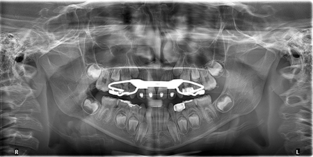
Fig. 6A

Fig. 6B

Case Study #3
A 9-year-old female presents with a Class I malocclusion with severe crowding. The panorex shows very impacted canines and crowding. (Figs. 7A, 8B) The treatment plan was a phased treatment with an expansion appliance and 4 anterior brackets. The outcome is more space for the canines. (Figs. 7B, 8B)
Fig. 7A

Fig. 7B

Fig. 8A

Fig. 8B

Is there evidence in the literature that supports this treatment modality?
The systematic reviews suggest that RPE appliances appear to improve the position of impacted maxillary canines and increase the likelihood of spontaneous eruption during mixed and early permanent dentition3,4. The use of an RPE has allowed 65% to 80% of the palatally displaced canines to erupt while, without the use of RPE the chance of eruption drops to 10%-13%4. The success rate of canine eruptions following an RPE treatment, with or without additional appliances, ranged between 65.7% and 85.7%. The success rate appeared to be higher when compared to the extraction of primary canines alone, where the rate ranged between 50%-60%.3
When is the best time to use expansion appliances to help reduce the chances of impacted canines?
A higher success rate of RPE treatment in younger children may be attributed to an early diagnosis of impaction. Therefore, establishing an early diagnosis of impaction is crucial to increase the chance of spontaneous eruption of canines. Studies have reported that the prognosis of successful spontaneous eruption of impacted canines worsens with increasing age7,14. Consequently, the patient should be diagnosed in mixed dentition, well before the eruption of the maxillary canines (mean eruption age is 12.3 years old for females and 13.1 years old for males)11. A panoramic radiograph is the most popular evaluation method identified in all studies and systematic reviews. It is also reported that the detection of an impacted maxillary canine from a panoramic radiograph could be made as early as 8 years age12.
So should you expand? You Definitely Should……
Oral Health welcomes this original article.
References
- Bedoya MM, Park JH. A review of the diagnosis and management of impacted maxillary canines. J Am Dent Assoc. 2009 Dec;140(12):1485-93.
- Bishara SE. Impacted maxillary canines: a review. Am J Orthod Dentofacial Orthop. 1992;101:159–71.
- Boonpratham, Supatchai, Pariyatdulapak, Natnicha, Poonpiriya, Thongchai, Peanchitlertkajorn, Supakit and Saengfai, Nuntinee Nanthavanich. “The efficacy of rapid palatal expansion on the eruption of impacted maxillary canine: a systematic review” Australasian Orthodontic Journal, vol.37, no.2, 2021, pp.273-283.
- De Stefani A, Bruno G, Visentin S, Lucchi P, Gracco A. Rapid maxillary expansion for interceptive orthodontic treatment of palatally displaced canine: A systematic review. Eur J Paediatr Dent. 2021 Jun;22(2): 139-143.
- Ericson S, Kurol J. Radiographic examination of ectopically erupting maxillary canines. Am J Orthod Dentofacial Orthop. 1987 Jun;91(6): 483-92.
- Ericson S, Kurol PJ. Resorption of incisors after ectopic eruption of maxillary canines: a CT study. Angle Orthod. 2000 Dec;70(6):415-23.
- Falahat B, Ericson S, Mak D’Amico R, Bjerklin K. Incisor root resorption due to ectopic maxillary canines: a long-term radiographic follow-up. Angle Orthod. 2008 Sep;78(5):778-85.
- Grisar K, Denoiseux B, Martin C, Hoppenreijs T, Calburean F, Politis C, Jacobs R. Treatment for critically impacted maxillary canines: Clinical versus scientific evidence – A systematic review. J Stomatol Oral Maxillofac Surg. 2022 Jun;123(3):e12-e19.
- Kaczor-Urbanowicz K, Zadurska, M, Czochrowska, E, Impacted Teeth: An Interdisciplinary Perspective. Adv Clin Exp Med 2016, 25, 3, 575-585.
- Kokich VG. Surgical and orthodontic management of. impacted maxillary canines. Am J Orthod Dentofacial Orthop. 2004;126:278-83.
- Litsas G, Acar A. A review of early displaced maxillary canines: etiology, diagnosis and interceptive treatment. Open Dent J. 2011 Mar 16;5:39-47.
- Sajnani AK, King NM. Early prediction of maxillary canine impaction from panoramic radiographs. Am J Orthod Dentofacial Orthop. 2012 Jul;142(1):45-51.
- Stivaros N, Mandall NA. Radiographic factors affecting the management of impacted upper permanent canines. J Orthod. 2000 Jun;27(2):169-73.
- Strbac GD, Foltin A, Gahleitner A, Bantleon HP, Watzek G, Bernhart T. The prevalence of root resorption of maxillary incisors caused by impacted maxillary canines. Clin Oral Investig. 2013 Mar;17(2):553-64.
- Topkara A, Sari Z. Impacted teeth in a turkish orthodontic patient population: prevalence, distribution and relationship with dental arch characteristics. Eur J Paediatr Dent. 2012 Dec;13(4):311-6.
About the Authors

Dr. Pinar Alkumru received her DDS from Istanbul University, her MSc and PhD from Ankara University and her DSAT from University of Toronto. Dr Alkumru teaches at University of Toronto since 2011, and owns a private clinic in Istanbul.

Dr. Pavel Sectakof received his DDS Dip Ortho and Msc degrees from University of Toronto. Dr Sectakof is an assistant professor at University of Toronto since 1993 and has two clinics in GTA area.
RELATED ARTICLE: Management of a 12-Year-Old-Male with a Class III Malocclusion and Canine Impaction


