
Introduction
Class II subdivision malocclusions can be difficult to treat because of their asymmetric occlusal relationships. An accurate diagnosis, etiologically based when possible, is essential to perform a correct and efficient orthodontic treatment.
The aim of this article is to illustrate a case report treated with customized lingual brackets in association with a simple vestibular device.
What is a Class II subdivision? Angle1 described it as a separate group of Class II malocclusions characterized by a unilateral Class II molar relationship. Statistically they make up around 50% of all Class II malocclusions and asymmetries. The main reason for these corrections is due to the functional implications.
Their aetiology could be dentally related, skeletally related, or a combination of both2. Orthodontic researchers and educators have discussed this definition a lot (even too much) and they have divided this specific malocclusion into type 1 and 2.
In type 1 the mandibular first molar has a distal position on Class II side and coincidence of the maxillary dental midline with the facial midline and deviation of the mandibular midline were observed.
In type 2 the maxillary first molar on the Class II side has a mesial position and coincidence of mandibular dental midline with the facial midline and deviation of the maxillary midline were observed3.
An optimal orthodontic treatment is the one that solves the entire list of problems with minimum discomfort and risk (including decalcifications) for the patient in the easiest way for the clinicians.
Starting from the consideration that Class II subdivision type 2 malocclusions very often have a mesio-rotational problem of the maxillary first molar, we find Carriere Motion device to be very practical and effective in the clinical usage.
This device consists of mold injected, nickel free stainless steel arm running from the maxillary cuspid to the first molar. Since it has a half sphere socket design on the molar pad it allows an easy rotation of the molar. The cuspid pad has a hook for an elastic going to a vestibular first lower molar button bracket.
Our practices located in Italy are only using customized lingual bracket for fixed orthodontic treatment because we want to provide the fastest treatment time, with the maximum tooth precision position, and with the minimal decalcification risk especially with adolescent patients.
Case Report
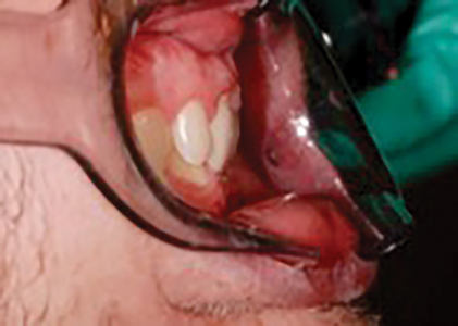
The patient presented with a Class II subdivision right type 2 malocclusion associated with a severe deep-bite and upper and lower crowding. He had functional limitations in the lateral movements of the mandible and frequent muscular tension headaches.
Pretreatment records
Fig. 1

Fig. 2

Fig. 3

Fig. 4

Fig. 5

Fig. 6

Fig. 7

Fig. 8

Fig. 9

Fig. 10

Fig. 11

Fig. 12
Pretreatment radiographs
Fig. 13
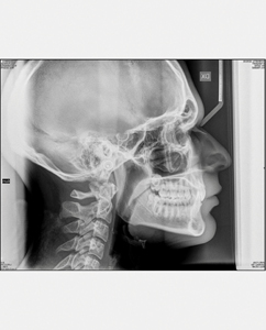
Fig. 14

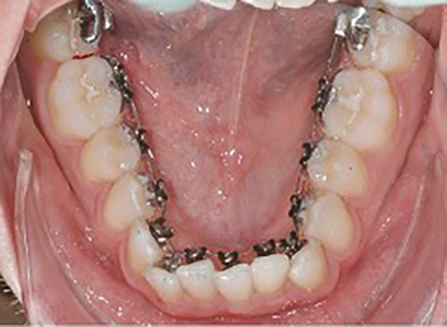
A fully customized lingual appliance (WIN) was installed and levelling and aligning started. At the second clinical visit two months later the patient reported that his headaches had been eliminated. This was likely due to a ‘splint effect’ from the lingual braces. (Figs. 15-16)
Treatment in progress
Fig. 15

Fig. 16
After 12 months, when a good levelling and intrusion was obtained we mounted the Carriere Motion appliance on the right side and a composite button on the lower first right molar. Elastics were prescribed. The patient was very cooperative wearing the elastics (3/16” 8 oz), removing them only for cleaning and eating. (Figs. 17-22)
Fig. 17
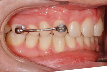
Fig. 18

Fig. 19

Fig. 20
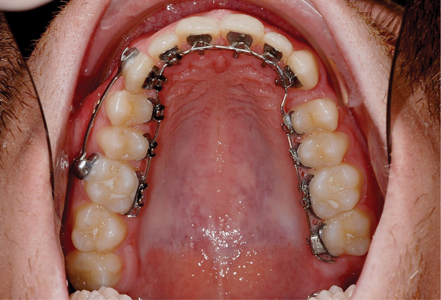
Fig. 21

Fig. 22

Six months later the carrier motion was removed when an over-corrected Class I was achieved. At that time, 018 x 18 TMA’s were placed and the finishing phase started.
The total treatment time was around two years and the torque control was superlative.
Both the gummy smile and the deep bite were reduced and a Class I occlusion was obtained bilaterally. (Figs. 23-36).
Posttreatment radiographs
Fig. 23
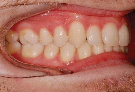
Fig. 24
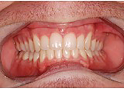
Fig. 25

Fig. 26
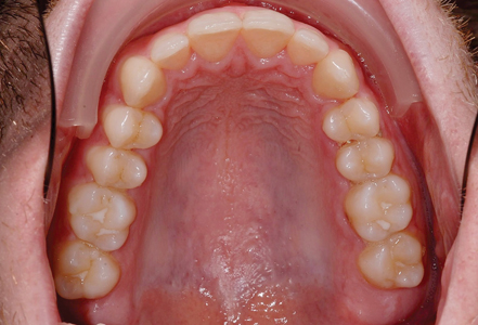
Fig. 27

Fig. 28

Fig. 29

Fig. 30

Fig. 31

Fig. 32

Fig. 33

Fig. 34
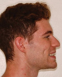
Fig. 35

Fig. 36

Conclusions
Customized Lingual Orthodontic treatments are now the golden standard of fixed orthodontics for their ability to reduce the risk of decalcification4, to obtain a very predictable tooth position, to reduce the treatment time5 while providing a truly invisible option.
Orthodontists who have tried non customized lingual treatments in the past probably would be impressed to realize how much the digital innovation has improved this technique that was born in the ‘80s.
Having the buccal side free from brackets has another advantage: the opportunity to use transparent composite buttons or devices like the carriere motion to easily distalize sectors keeping the full control of the movement of the teeth especially for patients that are afraid of mini screws or other TADs.
Oral Health welcomes this original article.
References
- Angle EH. Classification of malocclusion. Dent. Cosmos.. 1899;41: 350-75.
- Minich CM, Araújo EA, Behrents RG, Buschang PH, Tanaka OM, Kim KB. Evaluation of skeletal and dental asymmetries in Angle Class II subdivision malocclusions with cone-beam computed tomography. American Journal of Orthodontics and Dentofacial Orthopedics. 2013 Jul 1;144(1):57-66.
- Janson G, de Lima KJ, Woodside DG, Metaxas A, de Freitas MR, Henriques JF. Class II subdivision malocclusion types and evaluation of their asymmetries. American journal of orthodontics and dentofacial orthopedics. 2007 Jan 1;131(1):57-66.
- Wiechmann, Dirk, et al. Lingual appliances reduce the incidence of white spot lesions during orthodontic multibracket treatment. American Journal of Orthodontics and Dentofacial Orthopedics, 2015, 148.3: 414-422.
- KNÖSEL, Michael, et al. Lingual orthodontic treatment duration: performance of two different completely customized multi-bracket appliances (Incognito and WIN) in groups with different treatment complexities. Head & Face Medicine, 2014, 10.1: 1-12.
About the Author
 Gabriele Floria Certified Specialist in Orthodontics and Dentofacial Orthopaedics Lingual Orthodontic Clinical Instructor, Firenze Italy.
 Iris Floria Microbiology undergraduate student University of Glasgow, Glasgow, UK. The corresponding author’s address: gabriele@floria.it


