

The Role of Intra-oral Digital Scanning with Enhanced Process of Care
The addition of digital scanning to enhance clinical and technical procedures is a consideration for many dental practices. My training as a dental hygienist has taught me that the biggest benefits of digital intra-oral scanning present themselves in the overall patient experience, documentation, diagnosis, education and treatment planning. As clinicians we often focus on the clinical merits of introducing new technologies into our workflows. Another important aspect is to consider the positive patient experience that shows what we can do and enables them to become emotionally invested and motivated to follow through with treatment. There has never been a better time to offer and deliver comprehensive dental care. Advancements in research and development in all facets of dentistry have enabled patients to keep their teeth for a lifetime. In fact, science tells us that tooth wear may be considered a physiological process with an annual rate of wear of approximately 11μm.1 If extrapolated over a lifetime, the mathematics suggest it would take 100 years to lose 1mm of enamel. Coupled with increased consumer awareness regarding smile enhancements, the dental professional has substantially more merit to devise a customized prevention plan.
Dentistry can be more than a commodity of single dental procedures. As educators and advocates of oral health, we have an opportunity to advise patients of the best methods that will not only maintain the integrity of their teeth but will result in minimally invasive dentistry and overall improved oral and systemic health. The challenge to this is figuring out what form of communication can most efficiently enable patients to remember the discussion, value the advice, and more importantly, take actions towards optimizing their oral health.
I have found that implementing intraoral digital scanning into the dental hygiene process of care significantly enhances patient communication and enables the dental professional to share their concerns and intentions authentically. An intraoral digital scan can provide more nuanced information and serve as an adjunct to traditional charting, radiographs, and photographs. The patient experience is augmented since the process of capturing a scan is often faster and more comfortable than traditional impressions. With the iTero Element® 5D imaging system, the dental professional is equipped with several tools to confidently navigate real-time prevention plan options.
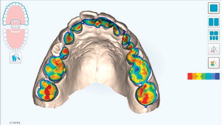
The iTero® Viewer and Occlusogram colour clearance map show the contact relationship between the upper and lower arches. In order to facilitate consistent assessments and patient messaging, I created a system based on the distribution of colour on the Occlusogram according to size, location, symmetry and intensity.
In the following example (Figure 1), the 3D model presented in the gallery Occlusogram view provides an instant relatable visual to identify areas of potential risk based on the display of colours The patient can easily recognize their dentition, and a high-level overview conversation about the general shape and form of the arches can begin. A narrowed arch form is identified suggesting an etiology for the crowding dentition. The lingually inclined posterior teeth suggest forces are not directed onto the occlusal surface along the axis of the tooth. Ideally, occlusal forces are transmitted vertically along the axis of the tooth and absorbed by the periodontium.2 (Figure 2). Vertical forces are less harmful because they provide axial stimulation to the teeth and bone while horizontal forces are extremely damaging via torqueing and off-loading.3 The elaborate distribution (size) of colour across the entire occlusal surface of the posterior teeth, ranging from 0.0mm to 1.0mm of clearance prompts the clinician to have increased awareness for the potential of interferences during function. Zero to minimal clearance (marked by red and orange colours) on non-functional cusps (location) may indicate premature wearing, chipping, receding soft tissue and abfraction lesions due to a potential increase in lateral and oblique forces . Lack of ‘symmetry’ from right to left or front to back prompt observations for unusual wear patterns and restoration integrity. In consideration of ‘intensity’, there is one rule, no ‘red’ on anterior teeth as they are not physiologically designed to endure the heavier contact reserved for posterior teeth.
Patients commonly enjoy the personalized interpretation of an Occlusogram analysis. I often give the analogy of a tarot card reading, informing the patient how the Occlusogram can identify areas of current and potential future concern. The benefit of this reading however is that our prescribed treatment plans can alter the outcome of the patients ‘cards’.
The Invisalign® Outcome Simulator tool can be utilized to help patients comprehend and visualize the goals of treatment. As clinicians we can use the imaging of the current dentition and simulated outcome to discuss our findings using the problem, consequence and solution model. By having a picture of the current dentition next to the simulated outcome, the projected changes and improvements to correct alignment, eliminate crowding and alleviate the pressure on the anterior teeth can be easily understood by the patient (Figure 3). The Invisalign Outcome Simulator tool can also be annotated with notes and shapes to draw attention to specific goals and assist with post-appointment consultation recall (Figure 4). It can be shared with the patient via email, allowing the patient to toggle between their current dentition and potential clinical result. The ability to view the simulation after the appointment can assist with communicating to family members the benefits of the proposed clear aligner therapy, allow patients to reflect on their dental visit, and perhaps even share it with others as a ‘wow’ experience.

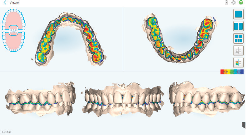
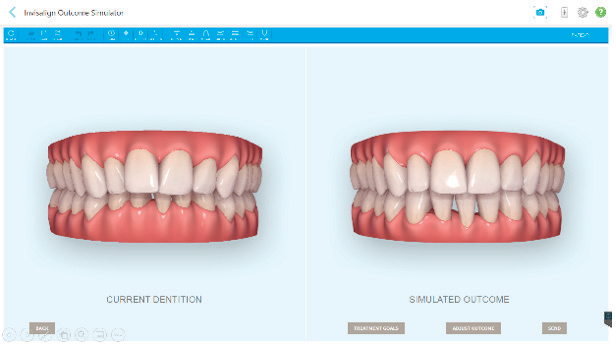
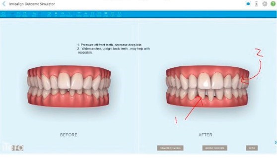
Figure 4
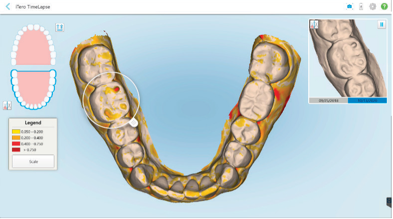
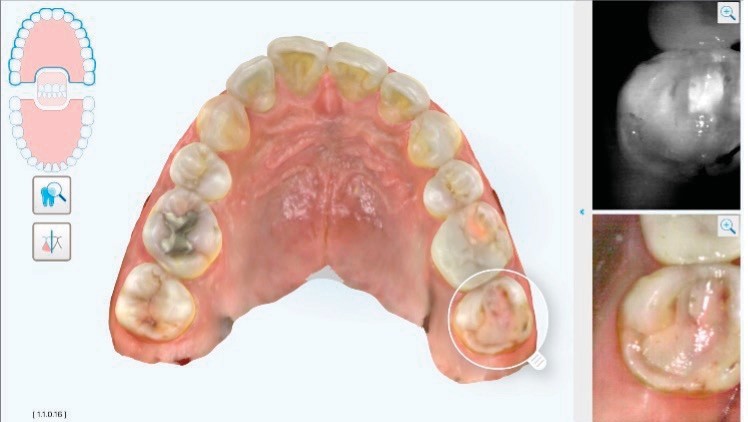
Daily trials of recording, analyzing, and evaluating minute oral changes test our abilities to be responsive and proactive in real-time. iTero® TimeLapse technology has become an analytic revolution for identifying and visualizing oral changes in soft tissue, hard tissue enamel and tooth movement in the range of 0.050mm to greater than 0.750mm! As a dental hygienist, the periodontal health of my patients is of utmost importance. In the past, I would utilize charting graphs to explain alterations in attachment levels (Figure 5). Although the graphics looked impressive, it was difficult for patients to navigate the location and interpret the multiple charting lines. As a clinician, these charting graphs are time consuming to analyze and evaluate over time. Now, using a stunning visual that compares historical scans to a current scan, I can superimpose scans, and let the software determine the degree of change. Patients can see for themselves the changes in their tooth wear, tooth movement, and changes in gingiva over time (Figure 6) and the close-up of the magnified field focuses on the area of concern (Figure 7).
We experience the same mesmerizing detail with hard tissue. In the following example, changes to the enamel surface can be seen due to an erosive process (Figures 8 and 9).
In both instances, the digital illustrations inspire us to be better oral detectives and clinicians. When considering? severity and the passage of time, we are prompted to ask more and better questions to determine the etiology of the deterioration. The data is available almost immediately upon completion of capturing the digital scan, creating opportunities to eliminate additional visits to review diagnostics, supporting real-time prescribed treatment or referrals, and more importantly to encourage a quality patient-led interaction.


The iTero Element 5D intraoral scanner is the first 3D scanner that integrates near-infrared imaging (NIRI) technology. It aids in the detection and monitoring of interproximal carious lesions above the gumline without using harmful radiation.4 The near infrared light transillumination presents low scattering and absorption of wavelengths allowing differentiation between healthy and carious tissue.5 Practitioners also have the ability to view multiple dimensions of data as well as virtually manipulate the model for a complete comprehensive view of the teeth, NIRI imaging, and intra-oral photos. Scanning with the iTero® Element 5D has eliminated the need for me to use other time-consuming imaging systems such as photography or an intra-oral camera. I have also found patients have a better understanding of the NIRI images, reducing their hesitation to proceed with prescribed radiographs to confirm suspected carious lesions.
In this example of a 5D scan, the upper arch is depicted with emphasis on tooth #2.7 indicated by the position of the magnifying glass. The bright white area along the mesial marginal ridge suggests an interproximal carious lesion may be present. The intra-oral photo illustrates loss of marginal integrity and less than ideal clinical appearance. (Figure 10).
Once again, the patient can readily identify the arch, the area where the tooth is located, and the NIRI and intra-oral image creates immediate interest in the oral condition. When coupled with the iTero® Occlusogram feature identifying localized increased occlusal contact on tooth #2.7 (Figure 10), an augmented diagnostic decision can be made for an excellent restorative outcome to protect and seal the tooth long-term.
Trust has always been imperative in the value exchange between the dentist, team members and patient. To increase consumer trust, dental practices must openly encourage dialogue, acknowledge patient needs, and personalize the education experience. The thoughtful incorporation and utilization of the iTero® Element intra- oral digital scanner enables precise diagnosis, and subsequently, predictable and effective therapies valued by patients. We the potential to transform our dental practices with scan-guided conversations, heightened clinical awareness and emotionally invested patients. I could not imagine practicing clinical dental hygiene any other way!
- Cuhna-Cruz J, Pahova H, Packard JD, Zhou L, Hilton Thomas Tooth wear: prevalence and associated factors in general practice patients. Community Dent Oral Epidemiol. 2010 Jun; 38(3): 228-234.
- Dicke, Non-carious Class V Lesions: What’s really going on? Access, November 2012
- McCoy G. “The Etiology of Gingival Erosion”. J Oral Impanto. 1982
- Data on file at Align Technology, INC. as of December 28, 2018
- Maria Melo, Augustin Pascual, Isabel Camps, Fadi Ata-Ali & Javier Ata-Ali. Combined Near-Infrared Light Transillumination and Direct Digital Radiography Increases Diagnostic in Approximal Scientific Reports (2019)9:14224
Ljiljana Hinton
 Ljiljana Hinton RRDH has over two decades of clinical Restorative Dental Hygiene experience. Her commitment to continued studies in Periodontics, Orthodontics and Restorative have confirmed that education is the most powerful resource for dental clinicians to facilitate patient comprehension with the goal of recognizing solutions to create optimal personalized oral health outcomes. After nearly three decades of working clinically full-time, Ljiljana joins Align Technology as a Clinical Digital Integration Specialist working with dental practices on the implementation of the iTero® Element digital workflow. Ljiljana finds professional and personal reward when invited to mentor, collaborate, train, and empower dental professionals to deliver science-driven, optimal standards of dental care.


