
Chief Complaint
A 35-year-old female patient was referred to our office by her husband on an emergency basis. The veneer on the upper left first premolar had fractured off. At that time all remaining residual cement was removed and the veneer sent to our lab to be cleaned and was silanated.
It was subsequently rebonded into position using Variolink. Some weeks later, she presented with a veneer on the lower right lateral incisor that had also dislodged and which was subsequently rebonded. At that point, the patient expressed frustration with the retention of her veneers and stated that she was very self-conscious about the overall appearance of her teeth and smile. As a result, she found that she was reluctant to smile and had become quite reserved and introverted. She was always apprehensive that her veneers would “fall off”.
Approximately two years prior to coming to our office, she explained that she had had a full mouth reconstruction employing a combination of porcelain labial veneers, crowns, and one implant.
The patient had an above average dental IQ and was very well informed about current trends in dental prosthetics. She brought up issues such as colour, shape, size, and thickness of the teeth, as well as facial structure, bite and gingival health.
The patient expressed great dissatisfaction with her current reconstruction, which she said, “did not live up to her expectations.” Her main concerns were:
1. She habitually felt a need to posture her lower jaw forward in order to improve her facial profile and to compensate for the appearance of sunken cheeks.
2. She said that prior to cementation of the case, she was never given the opportunity to visualize the final veneers and critique their appearance. Had she had the opportunity at try-in, she would have chosen a much lighter shade and veneers that were smaller and less square.
3. Another concern was with respect to the inflammation and profuse bleeding of her gums while brushing, flossing and eating such foods as apples.
Medical History
The patient is a 35-year-old female in good health. Her medical history was non-contributory. There were no systemic or local contraindications to the proposed treatment plan.
History of Present Condition
The patient had extensive crown and bridge performed in the maxillary and mandibular arches in addition to one implant placed at site 46. There were porcelain labial veneers placed from 15 to 25 and 35 to 45. The existing crown and bridge is approximately two and a half years old.
Upon closure, she reported that the teeth did not meet evenly and it was uncomfortable for her to occlude. She felt inclined to position her lower jaw forward in order to assume a more comfortable position. As well, she complained about a lisp when enunciating “s and “th” sounds.
Fig. 1
Hypermentalis, incompetent lip posture.

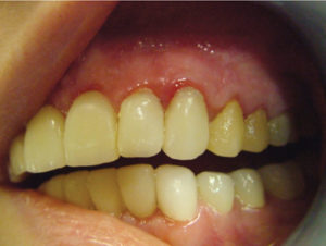
Fig. 2
Gingival inflammation due to impingement of crown margins into biological width.

Fig. 3
Poor plaque control and impingement of biologic width.
Oral Examination
A complete clinical and radiographic assessment of the hard tissue and soft tissues was performed. We discussed her occlusion and excursive movements. For all cases requiring comprehensive rehabilitation, patients are routinely referred for periodontal evaluation prior to commencing treatment. The gingival tissues were inflamed, edematous and bled easily upon light probing. This was due to the fact that the crown margins were overextended and impinging upon the biologic width. 1
Records were taken including diagnostic models, and intra and extra oral photos, comprising full face, smile line-relaxed and retracted together with right and left retracted smile. We e-mailed the photographs to our lab in order to communicate these concerns. Later, she would attend the lab so they could more accurately assess her facial contours and for shade selection. 2
Examination and Evaluation
The patient reported that her cheeks appeared sunken after the placement of the original crowns. During the original treatment, it was hypothesized that the vertical dimension had been increased perhaps more than was desirable, resulting in the appearance of sunken cheeks. It was determined, however, that she was overclosed and had an extremely deep overbite. As well, there was excessive gingival display about the maxillary anterior teeth resulting in a “gummy smile”. The combination of the above two factors with reduced vertical dimension and excessive gingival display, resulted in a dominant toothy appearance. 3
Treatment Plan
The patient underwent an initial sanative phase of periodontal scaling and root planing. She was prescribed Peridex™ rinses twice daily in conjunction with an electric toothbrush and unwaxed floss. To reduce the excessive gingival display and create more harmonious gingival contours, an esthetic crown lengthening procedure was planned. 4 This would also serve to restore the dimension for the development of a healthy connective tissue attachment thereby correcting the impingement of the biologic width.
Mockup
Due to the fact that the patient was dissatisfied with her aesthetics, phonetics, and facial contour, it was decided to arrange for the laboratory to prepare a detailed wax mockup from her study models. Once completed, this was presented to the patient for her evaluation. She was given an opportunity to critique the length and contours of the newly created dentition and once she was satisfied, we arranged for the lab to fabricate lab processed provisional crowns. The previous crown and bridge was removed, and the underlying teeth were re-prepared so as to provide ideal contours and emergence profiles in the provisional restorations. A polyvinyl siloxane impression was taken of the newly prepared teeth and sent to the lab for pouring. A facebow and centric occlusion bite registration were taken and the models were mounted in a semi-adjustable articulator. The lab was asked to open the vertical dimension approximately 2 mm in order to compensate for her overclosed position. We would be testing the patient’s tolerance to this new position while she was wearing the provisionals.
As a result of the patient’s aesthetic concerns, the lab created a mock wax-up of the teeth to the desired shape and contour. We ensured that the newly proposed occlusion would be functionally acceptable. Once the patient had an opportunity to visualize the mock-up, we asked for her approval in writing. We arranged to have our laboratory fabricate a temporary restoration, replacing all the existing crowns on the basis of this diagnostic template. Once the acrylic veneers were fabricated, the patient could try them out and we would be in a position to evaluate the need for functional modifications. As well, there was an increase of the vertical dimension and the new temporaries would allow us to verify whether the patient could tolerate this new position. We had indicated to the laboratory the proposed contours of the gingiva that would result after the esthetic crown lengthening so that these margins could be reproduced in the temporary crowns. This permitted improved adaptation of the gingiva at the gingival crest during healing. 5
Fig. 4
Post-op six weeks after final crown cementation. Note significantly improved gingival tone and stippling.

Fig. 5
Excellent gingival tone. Note stippling.

Fig. 6
Final result.

Periodontal surgery was accomplished using a surgical guide that mimicked the apical margins of the proposed new maxillary restorations. The surgical site was extended to the second premolars to maintain confluent gingival contours. A full thickness buccal flap was raised and osseous recontouring accomplished to mimic the final soft tissue contour and permit the development of a healthy attachment apparatus. A similar procedure was performed about the lower anterior teeth to create more harmonious contours and provide more dominance to the lower cuspids. The patient had reported that a previous attempt to lengthen her anterior teeth had been limited to the soft tissue. However, without reducing the underlying boney architecture, such efforts will result in soft tissue rebound and inflammation if there is encroachment upon the biologic width. ‘The bone sets the tone’ and must be addressed to ensure a stable soft tissue result.
We allowed the patient to wear this temporary for four to five months. During this period, she returned periodically to monitor healing, gingival adaptation, and maturation of the maxillary anterior segment. Once the functionality and esthetic requirements were achieved with the transitional restorations, maxillary and mandibular alginate impressions were taken to capture the final result. At that point final PVS impressions were taken of the prepared teeth. The lab fabricated temporary crowns for the prepared teeth using heat cured acrylic. These were subsequently cemented with TempBond®.
Final Crowns
Our laboratory fabricated PJCs for teeth #15-25 and PLVs for #11 and 21. In the mandibular arch, PJCs were fabricated for #33-43 and the PFM crown on the implant at #46. The decision was made to use the IPS E-max® system from Ivoclar Vivident® in order to achieve the desired aesthetics. These were tried in and we verified marginal fit, contact points and occlusion. Any adjustments were made accordingly and the case was cemented temporarily to give the patient a final opportunity for evaluation. Once she was satisfied and content with the final result, she gave her approval and signed in writing.
The preparation surfaces were cleaned with pumice paste on a prophy cup. The enamel was etched for 30 seconds using 30% phosphoric acid and the dentin was conditioned together with the enamel portions. Once these were rinsed off, the etching was verified and silane was applied. Then the dentin adhesive was carefully brushed onto the conditioned tooth surface. Final seating of the ceramic crowns was performed with low viscosity luting composite. Gentle pressure was applied until the restoration was placed in the correct position. Then any excess material was removed with a brush from the margins.
The restorations were tack-bonded into position with a Sapphire® light for only a couple of seconds. The interproximal areas were gently flossed with waxed floss to remove any excess cement and the floss was removed by sliding it out and not through the contact point in order to not cause any shift of the restoration.
These restorations were completely polymerized for 60 seconds from all aspects. The margins were finished using fine diamonds, Sof-lex® discs and Enhance® polishers. A preliminary check of the occlusion was made so that any gross discrepancies could be removed. The patient was permitted to try out her new dentition and two days later returned for fine occlusal equilibration in all excursions. Once the occlusion was finalized, a Brux-eze® appliance was fabricated which the patient was to wear nightly.
A follow up appointment was arranged approximately two weeks later in order to check the occlusion and gingival response. Both were excellent. The gingival was found to be pink, fibrotic and stippled. The patient reported that there was no bleeding upon brushing and flossing in any area of her mouth. 6
The above patient presented to our office with very high aesthetic expectations. Therefore, we felt it necessary to involve the dental technician and ask the patient to attend the laboratory in order to analyse the situation for herself and create a plan. It was felt that this method would lead more confidently to a predictable outcome. Thus, the final objectives were clearly defined between the dentist and technician as a result of thorough communication with the patient. This close communication from the inception enabled us to achieve a more predictable outcome. 7
Final Interview
Two months post insertion; we arranged a follow up appointment with the patient for the sole purpose of evaluating her appearance and perception of the final outcome. 8
The patient expressed that she felt much more confident because her looks had improved significantly. She noted that her pronunciation was much more distinct and she no longer had problems with “s” and “th” sounds. Friends and relatives have expressed that she has a more natural look now. She finds that she has a greater ability to focus and concentrate, as she does not feel distracted about her looks. She says that now she tends to be the one to initiate social interactions whereas before she found herself to be much more introverted. The patient’s psychological outlook has changed dramatically. She appears to be more energetic with an improved feeling of wellbeing. OH
Oral Health welcomes this original article.
Acknowledgement
We would like to acknowledge the contributions made by Dr. Alan Ghent to this article, as well as performing the periodontal surgical procedures associated with this case.
References
1. Success Is: “A Team Approach” Contemporary Maxillary Anterior Dental Implant Reconstruction. Dr Gary Brousell et al. Spectrum Dialogue, January 2012, Vol. 11 No. 1, 29.
2. Complex Full-mouth Rehabilitation Using All-Ceramics – Part 2. Dr Daniel Edelhoff et al. Spectrum Dialogue, March 2011, Vol. 10 No. 3, 38.
3. The Perio-Esthetic-Restorative Approach for Anterior Rehabilitation. Tomar N et al. J Indian Soc Periodontology. July 2013, Vol. 17 No. 4, 535.
4. The Effect of Esthetic Crown Lengthening on Perceptions of a Patient’s Attractiveness, Friendliness, Trustworthiness, Intelligence, and Self-Confidence. Dr Sam Malkinson et al. Journal of Periodontology, August 2013, Vol. 84, No. 8, 1126.
5. Complex Full-mouth Rehabilitation Using All-Ceramics – Part 2. Dr Daniel Edelhoff et al. Spectrum Dialogue, March 2011, Vol. 10 No. 3, 26.
6. Complex Full-mouth Rehabilitation Using All-Ceramics – Part 2. Dr Daniel Edelhoff et al. Spectrum Dialogue, March 2011, Vol. 10 No. 3, 23.
7. Complex Full-mouth Rehabilitation Using All-Ceramics – Part 2. Dr Daniel Edelhoff et al. Spectrum Dialogue, March 2011, Vol. 10 No. 3, 25.
8. Success Is: “A Team Approach” Contemporary Maxillary Anterior Dental Implant Reconstruction. Dr Gary Brousell et al. Spectrum Dialogue, January 2012, Vol. 11 No. 1, 40.
About the Author
 Dr. Tuvel maintains a private practice dedicated to general cosmetic and complex restorative dentistry in downtown Toronto, Ontario. He graduated from the University of Western Ontario Faculty of Dentistry in 1978 and has been in practice for over 30 years. He is a member of several dental organizations including the American Academy of Osseointegration and is certified in moderate sedation. Email: drtuvel@dentistryonbay.com.
RELATED ARTICLE: Treatment Planning: The Full Mouth Reconstruction
Follow the Oral Health Group on Facebook, Instagram, Twitter and LinkedIn for the latest updates on news, clinical articles, practice management and more!


