
Dental agenesis, the congenital absence of one or more teeth, is the most common developmental anomaly in humans and appears in 2.2-10% of the general population.1 The majority of cases are genetic in origin. In literature, congenital tooth agenesis can be categorized as anodontia, hypodontia, or oligodontia. Anodontia, the complete absence of teeth, is the rarest of the three categories. Hypodontia, refers to the absence of one to six teeth. Oligodontia is defined by the congenital absence of six or more permanent teeth, excluding third molars.1,2
The most affected teeth are maxillary lateral incisors, maxillary and mandibular second premolars, and the least affected teeth are maxillary central incisors, maxillary first molars, and mandibular canines. Several studies have reported that bilateral agenesis of teeth is more common than the unilateral form.3,4
Oligodontia is a rare condition that can occur in association with genetic syndromes, or as a non-syndromic isolated familial trait, or as a sporadic finding. The prevalence of non-syndromic oligodontia varies from 0.16% to 0.36% depending on the population studied.2,5,6 Oligodontia in non-syndromic form or familial form is more common than syndromic form. A thorough physical exam of the hairs, nails, sweat glands, and eyes to check for congenital disorders can be conducted to differentiate between syndromic and non-syndromic oligodontia. Oligodontia patients often display characteristics such as conical shaped teeth, microdontia, delayed eruption of permanent teeth, an increased freeway space and retained deciduous teeth. In addition to the loss of function and esthetic compromise, psychosocial development is an important concern in the oral rehabilitation of growing patients.
The biologic basis of congentially absent permanent teeth is explained partially by the failure of lingual or distal proliferation of tooth bud cells from the dental lamina. Tooth agenesis can be caused by environmental factors such as viral disease during pregnancy, metabolic imbalances, irradiation, tumour, or due to genetic factors, or to both. Mutations in transcription factors MSX1 and PAX9 have been identified as playing a major role in autosomal dominant oligodontia. Both genes play a critical role in early tooth development. All mutations of the PAX9 gene identified to date have been associated with non-syndromic form of tooth agenesis.7
Oligodontia patients are faced with significant dental concerns requiring a multi-disciplinary approach, in treatment management, involving a team of dental specialists. In Ontario, young oligodontia patients are usually identified by their general or pediatric dentist and can be referred to The Ontario Cleft Lip and Palate/Craniofacial Dental Program. This program is funded by the Ministry of Health and Long-Term Care and provides financial assistance for the specialized dental needs of affected individuals. The eligible patients who are accepted into the program are entitled to one course of treatment, completed by 22 years of age, as long as certified dental specialists complete the procedures. The patients do have a choice in the dental specialists performing the treatments, as the team of dental specialists involved does not have to be hospital based.
Case Report
A healthy, 19-year-old male patient presented to the office. As an oligodontia patient, he was funded in part by The Ontario Cleft Lip and Palate/Craniofacial Dental Program. Dr. Yair Lenga was the periodontist involved in his care. His past medical history was non-contributory. His family history revealed that he has a younger sibling, but no one in his family have congenitally missing teeth. Patient had orthodontic treatment as a child. He had been wearing the same set of Hawley retainers with pontics in place of the missing teeth for the past four years and the retainers no longer fit very well.
Patient expressed frustration towards having multiple missing teeth and retained deciduous teeth. He had just completed his first year of college, was shy and very uncomfortable with his appearance.
Extraoral examination revealed no abnormalities. Since patient had never reported any issues with hair, nails, eyes, and perspiration was normal, he was identified as being affected by non-syndromic form of oligodontia. Esthetic evaluation revealed the missing teeth in view when the patient smiled (Fig. 1). The maxillary midline was coincident with the facial midline. The occlusal plane was slightly canted upwards in the second quadrant.
Fig. 1A

Intraoral and radiographic examinations showed the following: slight conical appearance of maxillary central incisors, length discrepancy between the maxillary central incisors, peg-shaped lateral incisor, mismatched maxillary and mandibular dental midlines, missing permanent teeth (#13, 23, 24, 41, 31, 45, and 27; as well as #18, 38 and 48), the coronal structure of #28 was formed and visible on the panoramic x-ray but remained unerupted, #85 deciduous molar remained in the space of the missing #45, #71 and 81 mandibular deciduous central incisors remained in the area of the missing permanent central incisors, the roots of #12 and 14 appeared to be converging. The teeth #43 and 44 had supra-erupted into the opposing edentulous area, further accentuating the upward cant of the occlusal plane in this area. There was generalized gingival inflammation as the patient’s oral hygiene habit was not great and required improvement (Figs. 2-5). The occlusal views of the maxillary and mandibular arch showed lack of buccal and lingual width of the dentoalveolar ridge in areas of tooth agenesis. This deficiency in ridge width was especially prominent in the area of the mandibular central incisors although the bone height appeared to match that of the adjacent permanent teeth in this area (Fig. 6).
Fig. 2

Fig. 3

Fig. 4
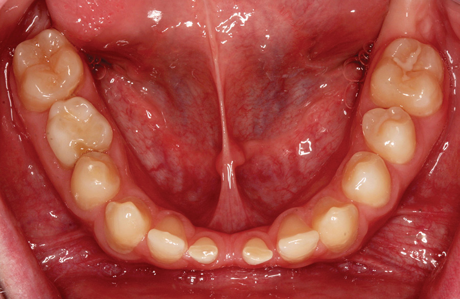
Fig. 5

Fig. 6

A few treatment options were presented and discussed with the patient and the parents after the initial evaluation. Even though the patient was already considered an adult and could legally make his own decisions, he wished for his parents to be included in the discussions as they had been involved with his treatment over the years and were financially responsible for the treatment. The retained deciduous teeth #71, 81, 85 were to be extracted, during healing, pontics were to be added to the existing Hawley retainer to replace the extracted teeth. The following treatment plans were presented: 1. A combination of fixed partial denture at #14-X-12, with implant restorations at #23, 24, 31, 41, and 45; 2. Conventional removable partial dentures for both the maxilla and the mandible. The mismatch in lengths of the maxillary central incisors and slight conical shapes of both sets of the maxillary central and lateral incisors had also been brought to the patient’s and his parents’ attention. The patient expressed no concerns with the appearance of the maxillary incisors and wished only to restore the missing permanent dentition.
The patient, being so young, rejected the removable dental prostheses option and chose to have fixed dental prostheses as the long-term restorative solution. One major concern with placing dental implants for a patient at this age was the possibility of the implants, along with the implant restorations, becoming submerged over time as the patient continues to grow. This was discussed with the patient and delaying the surgery for implant placement was presented as an option. However, the patient and his parents were eager to proceed and were willing to accept the risks associated with having implants placed before the growth was completed. To mitigate this risk, the patient was referred to his physician for a wrist X-ray to confirm the status of skeletal maturation prior to treatment.
Prior to beginning any prosthodontic treatment, carefully planning was always indicated. For this patient, preliminary alginate impressions were made and diagnostic casts poured and mounted on a semi-adjustable articulator. Diagnostic waxing was completed as part of the preoperative planning (Fig. 7). This allowed the patient to envision the final outcome of the treatment as planned prior to initiating any irreversible treatment. This also helped in managing a patient’s expectations when it comes to limitations one may face with restoring a case. In this patient’s case, this allowed for him and his parents to see the desired shape and positions of the planned restorations along with the existing maxillary anterior teeth in their unaltered shape. The wax-up also demonstrated to the patient the expected esthetic compromise in the edentulous areas where the ridge is atrophic, as well as the maintained occlusal plane can’t with the planned restorations in place as the restorative space is limited by the supra-eruption of the teeth in the third quadrant. Patient confirmed that he was satisfied with the restorations as planned without correcting any of the imperfections of his existing dentition. Since the #22 would be unaltered, the anatomy of the existing #12 would be maintained when the retainer for the fixed dental prosthesis was being designed and fabricated.
Fig. 7

Cone beam CT scans of the maxilla and mandible were taken during the initial treatment planning phase. Both edentulous areas, quadrant 2 and mandibular anterior, were extremely atrophic, with the mandibular anterior region being most severe, with 2 mm of buccolingual width in most of the areas. Ridge augmentation procedures were performed in both areas by Dr. Lenga, the periodontist. At the time of ridge augmentation, #85 deciduous molar was extracted and #45 implant was placed. The patient and his parents were fully aware of the high risk of complications associated with the grafting procedure due to the complexity of the case. Updated cone beam CT scans were taken after maturation of the hard tissue grafts (Figs. 8 & 9). One can appreciate the thickness of the grafted bone over the native bone in the recipient sites, which was identifiable due to the different radiodensities displayed on the scan. The ridge augmentation procedure had increased the volume of the available in the maxilla substantially, though not in height. The width of the available bone at the mandibular anterior region had improved with the graft, but still presented with much limitation. The first 5 mm of the crestal ridge, as well as areas immediately surrounding the deciduous incisors, showed width of less than 5 mm. This presents substantial limitation for implant planning. Even with a narrow diameter implant, at 3 mm, the periodontist would still have to sacrifice some of the crestal bone height in order to engage the full length of implant in the available bone, at a much more apical position than if the ridge was wider at the crest. The implants would have no problem with osseointegration, but the more apical position of the implants and the inevitable bone reduction in the area would have an effect on the hard and soft tissue architecture in the area and would become noticeable.
Fig. 8

Fig. 9

By keeping in mind the available bone as shown on the CT scans, the following implants were selected and placed: AstraTech OsseoSpeed TX 3.5×11 mm implants for both #23 and 24; AstraTech OsseoSpeed TX 3.0x11mm implants for both #31 an 41; and AstraTech OsseoSpeed TX 5.0x9mm implant for #45. All implant sites healed without any complications. Once the osseointegration of the implants was confirmed by Dr. Lenga, the patient returned to the office to begin the restorative phase of his treatment. As predicted earlier at time of implant planning, all the surgical sites healed well but there was noticeable change of tissue architecture around the implants. By comparing the preoperative and postoperative intraoral pictures, one can appreciate this change in tissue architecture. This was an issue that we were able to foresee but unavoidable due to the complexity of the clinical situation (Fig. 10).
Fig. 10

Since the patient was ready for the restorative phase, #12 and 14 were prepared as retainers for fixed dental prosthesis. CAD/CAM gold-hue Titanium Atlantis abutments were designed and ordered for the implant restorations (Figs. 11A-B & 12). Custom abutments were designed to support the emergency profile, as well as provide adequate retention and resistance form of the abutment under the implant restoration. In the case of our oligodontia patient, because of the lack of soft and hard tissue architecture in the edentulous area around the implants, there was very limited control over the emergency profile of the implant abutment and restorations. The emergency profile is usually established with the abutment starting out at a narrower diameter, from the collar of the implant, extending out to a larger diameter, in the anatomic shape of the tooth being restored, just below the gingival margin. This transition, in diameter and shape, creates an illusion that the restoration is emerging from the ridge. For our patient, because of the lack of tissue in between the implants, in both the maxillary and mandibular implant sites, it was not possible to develop the ideal emergence profile with the custom abutments. This possible esthetic deficiency was discussed with the patient and his parents. A few options were presented. One option would be to splint the implants and use pink porcelain in the interproximal areas between the prosthetic crowns. Another option would be to leave the implant restorations unplinted and not use pink porcelain. The esthetics of the splinted restorations with pink porcelain would be superior, as it would give the lab an opportunity to help camouflage the soft tissue defect. However, patient’s oral hygiene was a concern. It is evident, from the chronically erythematous gingival tissues and inflammed interproximal papillae, that oral hygiene compliance was an issue even with frequent reminders throughout his treatment. The patient also expressed very little concerns about the esthetic outcome of the treatment. Therefore, a decision was made to restore the implants unsplinted, to aid in flossing and routine cleaning in between and around the implant restorations.
Fig. 11A

Fig. 11B

Fig. 12

Full contour zirconia restorations were designed, fabricated, and cemented onto the Atlantis abutments for the implants #23, 24, 31 and 41. Teeth #12 and 14 were prepared and full contour fixed dental prosthesis 12-X-14 was fabricated and cemented. As one can appreciate from the photo taken two weeks post-cementation (Fig. 13), the size and shape of the definitive restorations follow that of the diagnostic wax-up with a few minor corrections. The buccal contour of #14 was shaped to better follow that of the adjacent premolar, as the original contour of the natural tooth flared out buccally. As planned during the diagnostic wax-up phase, the slight upward cant of the occlusal plane was maintained, partly due to the limited occlusal space as a result of the supra-erupted dentition in quadrant three, which was left unaltered. One can also appreciate the esthetic limitation of the implant restorations in the #31, 41 interproximal area. As noted, patient’s oral hygiene habit was not great and despite the favourable soft tissue response around all the fixed dental prostheses, there was already obvious plaque accumulation around the base of the implant restorations at the two-week mark. This suggests that the decision to make the implant restorations as cleansible as possible was necessary.
Fig. 13

The post-insertion smile photo showed improved esthetics provided by the comprehensive dental rehabilitation, despite the anatomical challenges faced during the course of treat. The patient only had a limited display of the maxillary gingival areas while smiling and does not show the base of the mandibular incisors (Fig. 14). He was very happy with the functional and esthetic outcome of the rehabilitation. The complexity of this treatment was envisioned and properly managed for the patient. The restoration of esthetics and function had been achieved with an interdisciplinary team approach.
Fig. 14

For this patient, meticulous attention to the details required due to the complexity and limitations of the case, from diagnosis to fabrication of final restorations, allowed a controlled and logical treatment sequence. While many other prosthodontic management approaches have been reported, this case demonstrated one approach to comprehensive rehabilitation of a patient with oligodontia with successful results.
Oral Health welcomes this original article.
References
- Kotsiomiti E, Kassa D, Kapari D. Oligodontia and associated characteristics: assessment in view of prosthodontic rehabilitation. Eur J Prosthodont Restor Dent. 2007 Jun:15(2):55-60.
- Tavajohi-Kermani H, Kapur R, Sciote JJ. Tooth agenesis and craniofacial morphology in an orthodontic population. Am J Orthod Dentofacial Orthop. 2002;122:39–47.
- Silvameza R. Radiographic assessment of congenitally missing teeth in orthodontic patients. Int J Paediatr Dent. 2003;13:112-6.
- Kirzioglu Z, Koleser Sentut T, Ozay Erturk MS, Karayilmaz H. Clinical features of hypodontia and associated dental anomalies: A retrospective study. Oral Dis. 2005;22:399-404.
- Rolling S. Poulsen S. Oligodontia in Danish schoolchildren. Acta Odontol Scand. 2001 Apr:59(2):111-2.
- Polder BJ, Van’t Hof MA, Van der Linden FP, Kuijpers-Jagtman AM. A meta-analysis of the prevalence of dental agenesis of permanent teeth. Community Dent Oral Epidemiol. 2004 Jun;32(3):217-26.
- Guruprasad R, Nair PP, Hegde K, Singh M. Case report: nonsyndromic oligodontia. JIDA. 2011;3:450–454.
About the Author
 Dr. Ming-Yi Chou a fellow of the Royal College of Dentists of Canada in Prosthodontics. She completed her prosthodontic specialty training and obtained both a Certificate in Prosthodontics and Master of Science degree in Prosthodontics from the University of North Carolina, USA. Dr. Chou received her degree of Doctor of Dental Surgery from the University of Western Ontario, Canada, where she graduated with distinction in 2006. Dr. Chou is a member of the American College of Prosthodontists (ACP) and is currently the President of the Association of Prosthodontists of Ontario (APO). Dr. Chou lectures both locally and internationally, teaches at the Graduate Prosthodontics department at University of Toronto as an clinical instructor and has a private practice in Toronto.
RELATED ARTICLE: Transition From A Removable To A Fixed Implant-Supported Prosthesis: A Case Report


