
Abstract
Incorporation of dental implants to support fixed and removable prostheses is an effective treatment modality for edentulous patients that provides enhanced patient function, comfort and overall quality of life in comparison to a conventional complete removable prosthesis. Both fixed and removable implant-supported prostheses for edentulous patients have specific indications although in many clinical situations both prosthesis types are completely appropriate. The choice of the treatment modality is often ultimately made by the patient as a reconciliation of the patient’s treatment objectives with the willingness to undergo the corresponding treatment steps and the willingness to accept the fiscal commitments. Occasionally, patients rehabilitated with an implant-supported prosthesis may desire to transition to a different prosthesis type – either from an implant-supported removable to a fixed prosthesis or vice versa. Loss of implants, a desire for greater retention and a desire for ease of home care are three most common reasons that such transition from one implant-supported prosthesis type to another may be desired or required.
This case-report focuses on a transition of 75-year-old male from having an implant-tissue supported, bar-retained mandibular overdenture to having a full-arch fixed implant-supported prosthesis to address the patient’s concerns with comfort, retention and mastication. The patient’s history is significant for an earlier transition from a fixed implant-supported prosthesis to a removable one due to fracture of a critical number of implants.
This case report highlights the versatility of osseointegrated implants to achieve a variety of clinical objectives and the necessity of treatment planning to account for the possible future need to transition to a different implant-supported prosthesis type.
Introduction
An increase in the elderly population has resulted in an increase in demand for optimal management of completely edentulous patients. This patient population may have esthetic and functional demands which can be met with conventional removable prostheses as well as fixed and removable implant-supported prostheses. In the treatment of completely edentulous mandibles, implant-supported overdentures are becoming a frequent choice of treatment as they can address many complaints associated with conventional removable prostheses. Studies demonstrate very high success and survival rates for implants as well as implant-supported fixed and removable prostheses in edentulous mandibles [Kern et al. 2016].
Implant-supported overdentures offer much improved stability, function, mastication, proprioception, comfort, retention and quality of life in comparison to conventional removable prostheses. Many different types of attachment systems are available to retain implant-supported overdentures. The type of attachment may be chosen based on the desired degree of retention, anatomy of residual alveolar ridge, interocclusal space, hygiene access, removable prosthesis type, patient dexterity, financial considerations as well as number, position, and parallelism of the implants. Both splinted and non-splinted designs have been successfully used in anterior mandible, and the type of retention system used does not appear to have a significant impact on treatment success and patient satisfaction [Kim et al. 2010]. However, maintenance requirements may vary [Cehreli et al. 2010].
Prosthetic maintenance requirements for implant-supported overdentures range from simple repair or replacement of the attachment to remaking the prosthesis and are driven primarily by prosthesis design, the technique of connection to the implant, and the denture quality in terms of occlusal and ridge adaptation, material, and components. By contrast, implant number, implant type, and attachment type do not appear to be critical factors in determining prosthetic maintenance requirements [Assaf et al. 2017].
Multiple differences exist in the treatment planning and execution between removable and fixed implant-supported prostheses from a surgical and prosthodontic standpoint [Bolouri and Bhavani, 2014]. Four key differences are a need for lip support, the number and distribution of implants, vertical clearance, and precision of 3-dimensional implant placement. Arch-specific differences may also need to be considered. In general, lip support is more optimally provided by removable solutions. Three-dimensional implant positioning may be less critical with removable than fixed implant-supported prostheses. Fixed prostheses require more precise orientation, particularly if the implant abutments are expected to emerge along the long axes of the replacement teeth. Moreover, fewer implants are required with overdenture treatments. In the edentulous mandible, two implants are typically required for overdenture treatment while four to five implants are required for full-arch fixed implant-supported prostheses. Due to these differences between the two prosthesis types, not all clinical situations beginning with an implant-supported removable prosthesis can be readily transitioned to a fixed prosthesis.
Case Report
Chief Complaint and History of Chief Complaint
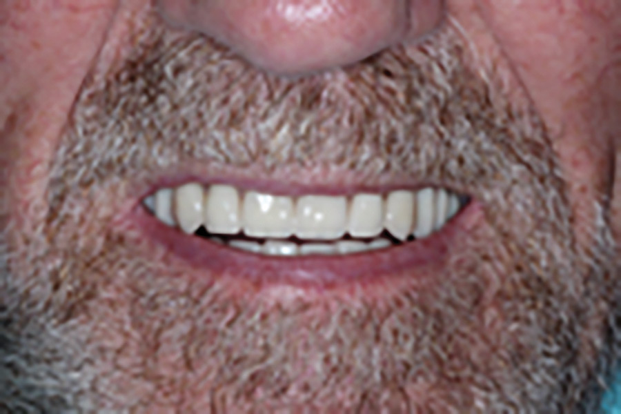
A 75-year-old male patient presented to the Graduate Prosthodontic Clinic at the University of Toronto, Faculty of Dentistry on an emergency visit with a chief complaint of a non-retentive mandibular implant-supported overdenture as well as secondary complaints of pain on chewing and poor aesthetics. The patient was completely edentulous; the maxilla was restored with a conventional complete removable dental prosthesis which was not aesthetically pleasing to the patient, and the mandible was restored with an implant-tissue supported bar-retained overdenture with supporting implants in the 32 and 42 positions. The implants (Branemark external hexagon, regular platform) were connected by a cast gold bar which engaged a corresponding clip in the intaglio surface of the mandibular overdenture (Fig. 1). The patient reported poor retention of the mandibular implant-supported overdenture and pain associated with the areas of the mental foramina bilaterally while chewing. The patient was not satisfied with the esthetics of the maxillary conventional complete removable dental prosthesis, mentioning that the anterior maxillary teeth were too long and too labially positioned.
Fig. 1A

Fig. 1B

Fig. 1C

The patient’s history was significant for being completely edentulous for over 50 years. The mandible had five interforaminal implants placed in 1987 to receive a full-arch fixed implant-supported prosthesis. After two years, the implant in the 44 site had fractured and the fixed mandibular prosthesis was modified to continue functioning on the four remaining implants. In 1990, implant in the 34 site fractured, and the patient was transitioned to an implant-tissue supported bar-retained overdenture on implants 32 and 42. A cover screw was placed on implant 41, and this implant was submerged.
Clinical and Radiographic Examination
Comprehensive extraoral and intraoral examination was performed. Findings upon extraoral examination were unremarkable except for slight loss of vertical dimension of occlusion which was noted based on clinical assessment of phonetics and esthetics and was alluded to by the visible wear of the prosthetic teeth on both prostheses.
Intraoral examination revealed that the maxillary prosthesis had insufficient retention despite evidence of multiple reline attempts to improve prosthesis retention and fit. Moreover, esthetics could be improved as the anterior teeth were set too labially. Mandibular implants had lingual inclination. The gold bar was screw-retained on the two implants and presented with minimal wear. The cover screw on the 41 implant was partially visible through the gingival tissue.
The mandibular prosthesis presented with suboptimal support, retention and stability and demonstrated minimal wear associated with the bar clip (Fig. 2). The radiographic examination demonstrated mild bone loss around the anterior mandibular implants, presence of the fractured implant in the 33 site and proximity of mental foramina to the crest of the ridge (Fig. 3).
Fig. 2A

Fig. 2B

Fig. 3

Management
Different treatment options were presented to the patient, and pros and cons of all the options were discussed. The patient opted for a new conventional complete removable dental prosthesis for the maxilla to address concerns with retention and esthetics and chose to transition to a full-arch fixed implant-supported prosthesis in the mandible. The latter was planned to include removal of the fractured implant in the 43 site, addition of two implants distal to 32 and 42, uncovery of implant 41, and fabrication of a fixed implant-supported prosthesis on five implants.
Important diagnostic factors were considered in the treatment-planning phase including the interocclusal space, need for lip support, and hygiene requirements. CT scan and diagnostic tooth set-up were obtained, and a surgical guide was fabricated for placement of mandibular implants in the 34 and 44 sites.
First, the fractured implant in the mandible was removed uneventfully (Fig. 4). Following adequate healing time of four months, implant 41 was uncovered and two implants (NobelReplace Conical Connection, 4.3 x 10 mm) were added in the 34 and 44 sites (Fig. 5). These implants were distally angulated to increase the anterior-posterior spread and to minimize the distal cantilevers of the planned transitional fixed prosthesis. Healing abutments were placed, and the overdenture was relieved, relined and re-inserted. During the healing time, a new vertical dimension of occlusion was established with the new maxillary conventional complete removable dental prosthesis which was fabricated to address the patient’s esthetic and functional concerns against the relined mandibular removable prosthesis (Fig. 6). After adequate healing of three months, the new implants 34 and 44 were successfully assessed for osseointegration.
Fig. 4

Fig. 5

Fig. 6A

Fig. 6B
Careful assessment of vertical clearance, implant angulation and position of the implant platform relative to the gingival margin was undertaken and informed the decision to restore distal-most implants 34 and 44 with 30 degrees angulated multi-unit abutments (Nobel Biocare, 30 degree Multi-unit Abutment Plus) and to restore the anterior three implants at implant-level by removal of existing standard abutments from 42 and 32 implants. An impression was obtained using appropriate impression copings in an open-tray manner and was used to fabricate a master cast (Fig. 7). The master cast was verified using intraoral splinting of non-engaging temporary cylinders, and an interocclusal record was obtained with a mandibular stabilized base plate and wax rim against the maxillary prosthesis at the optimal VDO. A mandibular tooth set-up was tried in, and adequacy of esthetics, phonetics and VDO were confirmed (Fig. 8). The metal framework for the mandibular prosthesis was designed, tried in and checked for passivity of fit. The definitive screw-retained prosthesis was fabricated and inserted (Fig. 9 & 10). The patient reported satisfaction with the provided care. The patient was re-assessed at six months post-insertion of the prosthesis and reported satisfaction with function and esthetics.
Fig. 7
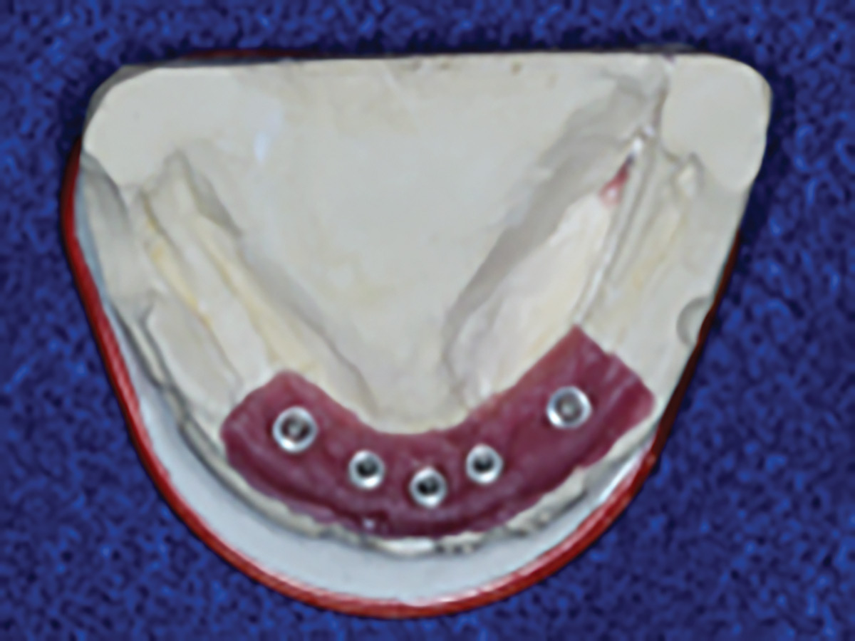
Fig. 8

Fig. 9A

Fig. 9B

Fig. 10

Discussion
Conventional mandibular complete removable dental prostheses may present with multiple challenges secondary to residual ridge resorption including reduced retention, instability of prostheses and soreness in the supporting mucosa. Rehabilitation of edentulous mandibles with residual ridge resorption has improved tremendously due to the emergence of implant dentistry [Goodacre & Goodacre 2017]. Despite the fact that implant-supported overdentures have resulted in improvement in stability, retention and esthetics, some edentulous patients may not be able to adapt to adequately fabricated conventional removable prostheses; hence, additional retention and support provided by fixed restorations may be desired. The patient presented in this case report had difficulty with an implant-tissue supported bar-retained mandibular overdenture and desired a fixed solution to replace his missing mandibular teeth.
Various elements may play a role in the retention of implant-supported overdentures. Chief among these are the quality of the removable prosthesis and its relationship to the supporting tissues and opposing occlusion, optimal location and fit of the attachment apparatus, and the location of the implants.
For the severely resorbed edentulous jaw, the all‐on‐4 concept, whereby two axial implants are placed in the anterior region and two angulated implants more posteriorly, has been utilized with success. This treatment modality minimizes the length of the cantilevers and results in a better load distribution [Patzelt et al. 2014].
Patients and clinicians may choose a removable or fixed implant-supported option based on various elements such as anatomic restrictions and cost considerations. Furthermore, motor difficulties often encountered with patients of advanced age may indicate a preference towards one option over the other [Assaf et al. 2018]. In the presented case, the patient desired the retention and stability and the resulting comfort associated with a fixed rehabilitation.
Both fixed and removable implant-supported therapies have been reported to be associated with high implant survival rates, patient satisfaction, and improved masticatory function. Similarly, both types of prostheses are impacted by prosthetic complications and the need for post-placement mechanical maintenance. However, implant-supported overdentures are reported to be associated with greater maintenance requirements. Although, implant-supported overdentures may be more cost-effective in the initial stages of care, higher maintenance fees may be encountered [Goodacre & Goodacre 2017].
It is crucial to note that each prosthesis type has advantages and shortcomings, and these need to be assessed on a case-by-case basis so that the treatment plan can be optimally customized to address each patient’s needs and desires.
Conclusion
This case report highlights the diagnosis and management of a completely edentulous patient with a poorly retentive suboptimal mandibular implant-supported overdenture and describes the process of its transition to a full-arch fixed implant-supported prosthesis. Implant treatment planning and placement must account for a possibility of future treatment evolution including a change from one prosthesis type to another.
Oral Health welcomes this original article.
References
- Assaf A, Daas M, Boittin A, Eid N, Postaire M. Prosthetic maintenance of different mandibular overdentures: A systematic review. J Prosthet Dent 2017;118:144-152
- Bolouri A, Bhavani V. Transition from a fixed implant dental prosthesis to an implant overdenture in an edentulous patient: A clinical report. J Prosthet Dent 2014;112:414-417
- Cehreli MC, Karasoy D, Kokat AM, Akca K, Eckert SE. Systematic review of prosthetic maintenance requirements for implant-supported overdentures. Int J Maxillofac Implants 2010;25(1):163-180
- Goodacre C, Goodacre B. Fixed vs. removable complete implant prostheses: A literature review of prosthodontic outcomes. Eur J Implantol 2017;10(1):13-34
- Kern JS, Kern T, Wolfart S, Heussen N. A systematic review and meta-analysis of removable and fixed implant-supported prostheses in edentulous jaws: Post-loading implant loss. Clin Oral Implants Res 2016;27(2):174-195
- Kim HY, Lee JY, Shin SW, Bryant SR. Attachment systems for mandibular implant overdentures: as systematic review. J Adv Prosthodont 2010;4(4):197-203
- Patzelt SB, Bahat O, Reynolds MA, Strub JR. The all-on-four treatment concept: a systematic review. Clin Implant Dent Relat Res 2014;16(6):836-855
About the Authors
![]() Elahe Behrooz obtained her first DDS degree in Iran and MBA degree from Carleton University. She obtained her second DDS degree with honours in 2016 and graduated from the graduate prosthodontics program at the Faculty of Dentistry, University of Toronto in September 2019. Dr. Behrooz can be reached at elahe.behrooz@mail.utoronto.ca.
Elahe Behrooz obtained her first DDS degree in Iran and MBA degree from Carleton University. She obtained her second DDS degree with honours in 2016 and graduated from the graduate prosthodontics program at the Faculty of Dentistry, University of Toronto in September 2019. Dr. Behrooz can be reached at elahe.behrooz@mail.utoronto.ca.
 David Chvartszaid is a dual specialist in Prosthodontics and Periodontics. He is the director of the Graduate Prosthodontics Program at the University of Toronto and the chief-of-dentistry at Baycrest Hospital. Dr. Chvartszaid can be reached at david.chvartszaid@dentistry.utoronto.ca.
RELATED ARTICLE: Full Arch Immediate Fixed Implant Rehabilitation: A Case Demonstration


