

Mirror-image twin is a unique phenomenon wherein the features on one identical (i.e., monozygotic) twin’s left side are mirrored on the other twin’s right side. For example, one twin may have a birthmark on their left cheek, while the other twin has a birthmark in the exact location but on the right cheek. Mirror twins occur when the fertilized egg splits after the lateralization begins. The prevalence of mirror twins can only be estimated from indirect evidence.1,2 Approximately 25% of monozygotic twins (n=75) in the United States3 and 23% of monozygotic twins (n=375) in Belgium4 had discordant handedness (one sibling is right-handed, the other is left-handed). However, handedness is determined mainly by the non-shared environment with fewer genetic influences.
While mirror twins share identical DNA, they may have distinct personalities, interests, and abilities, and they may also experience different health issues throughout their lives. Numerous studies have reported mirror-image dental anomalies in twins: aplasia,5 supernumerary teeth,6 or dental fusion.7 Contralateral craniofacial lesions such as cleft lip and palate,8 hemifacial microsomia,8 and unilateral condylar hyperplasia have been reported.9 The pathological phenomena suggest the importance of mirror-imaging.2
Recently, it has been demonstrated that palatal geometry measured on the intraoral scan can distinguish strangers and even identical twins with moderate certainty, facilitating human and victim identification.10 After selecting possible matches, an additional superimposition of the palatal scan can confirm the identity.11,12 However, recognizing a twin relationship might be concealed by the opposite laterality of siblings or twin relationship. Therefore, this study’s primary aim was to assess the effect of sagittal mirroring on the fit between twin siblings’ scans.
Compared to DNA analysis, the strength of the intraoral ante-mortem scan-based identification method is growing due to the rapid spread of intraoral scanning. However, the limitation is that dentists may focus on quadrant rehabilitation, limiting the scan to only one palate side. Similarly, a severe accident may damage the half palate. Although the palatal side-to-side asymmetry is relatively small,13 the total geometry and surface morphology may differ between sides. Therefore, the study’s secondary aim was to assess the fit between the mirrored scan and the original scan of the same individuals.
Material and Methods
Sample collection and preparation
A total of 174 participants were recruited through the National Twin Registry,14,15 61 monozygotic twin pairs (122 individuals) and 26 same-sex dizygotic twin pairs (52 individuals). All participants agreed to study participation by written informed consent. Ethical approval was obtained (36699-2/2018/EKU). Zygosity was determined using questionnaires.16 Evidence of previous orthodontic treatment was also recorded. Each participant’s palate was scanned three times using a Planmeca Emerald intraoral scanner (Planmeca Oy, Helsinki, Finland, version number Romexis 5.2.1) as previously described.11
All three scans of the 174 subjects (522 scans) were cropped, and teeth were removed in the GOM Inspect Suite software (GOM GmbH, Braunschweig, Germany). The cropped palatal model was duplicated, and one was mirrored in the y-axis (i.e., sagittal plane) (Figs. 1A and B). The palatal scans were aligned by the best-fit function using the iterative closest point algorithm.17 The mean absolute distance between aligned scans’ surfaces was calculated by dividing the integrated absolute distance by the area of the valid distance.
Fig. 1

Assessment of twin mirroring (between-siblings comparison)
The three scans of one sibling were superimposed with three scans of the other sibling (original group, nine superimpositions per subject). Additionally, the three original scans of one sibling were superimposed with three mirrored scans of the other sibling (mirror group, nine superimpositions per twin pair). A total of 2610 measurements were made.
Assessment of asymmetry (between-replicates comparison)
The original three scans within a subject were superimposed on each other (original group, three superimpositions). The three mirrored scans were superimposed with a non-mirrored replicate (mirror group, three superimpositions).
Statistics
The Wilcoxon test was used to compare the original and mirror mean absolute distance in the between-siblings and between-replicates comparisons. Mann-Whitney U test was used to compare the monozygotic with the dizygotic twins. The number of twin pairs in which the difference between the scans increased following mirroring was tabulated, as well as the number of pairs in which the difference decreased. Then the ratio of increased to decreased was calculated. The proportion in monozygotic and dizygotic was compared by Chi-square statistics. All analyses were made by IBM SPSS Statistics, Version 27 (Armonk, NY: IBM Corp., USA). A p-value less than 0.05 was considered statistically significant. The data in the text is given in the median and interquartile range.
Results
Assessment of twin mirroring between-siblings comparison)
The mean absolute distance between-siblings increased significantly (p<0.05) after mirroring in monozygotic twins from 0.428mm (0.317-0.510) to 0.440mm (0.339-0.507). (Fig. 2) There was no significant change in the dizygotic twins; 0.509mm (0.408-0.882) vs 0.497mm (0.377-0.807), p=0.149. The between-siblings mean absolute distance decreased in 27% of monozygotic twins and 22% of dizygotic twins. The percentage was not significantly different between the monozygotic and dizygotic groups (p=0.717). The mean absolute distance was significantly higher in dizygotic than monozygotic twins regardless of mirroring.
Fig. 2

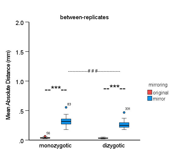
Assessment of asymmetry (between-replicates comparison)
The mean absolute distance significantly increased (p<0.001) in monozygotic twins from 0.036mm (0.030-0.043) to 0.316mm (0.270-0.358) and dizygotic twins from 0.034mm (0.028-0.039) to 0.249mm (0.216-0.303) after mirroring, (Fig. 3) but with the greater extent in monozygotic twins. The mean absolute distance was significantly higher in monozygotic than in dizygotic twins in the case of mirroring.
Fig. 3
Discussion
On average, the difference between the two siblings’ scans increased only by 12µm by mirroring one of the siblings’ palates. Nevertheless, the effect was negligible considering the discrimination potential (428-509µm) and the reproducibility of the scan (34-36µm), in agreement with previous findings.11 Furthermore, in 22-27% of the twins, the difference between scans decreased after mirroring, suggesting that these twins have a contralateral similarity. This prevalence resembles previous results.2,3,4 Although no data about their common chorion was available, those twins could be mirrored twins. There is evidence of mirror-image monozygotic twins, but only one study has described the same for dizygotic siblings.18 In a forensic investigation, making a mirrored alignment between two scans may have great value if the low deviation between scans indicates relatives. The mirroring process can confirm a possible twinning relationship and facilitate finding the victim’s family.
The dizygotic between-siblings values were higher, suggesting that the higher the 3D deviation between two persons’ scans, the higher distance in relatedness. Furthermore, the between-siblings values were much higher than the between-replicates ones, indicating that an intraoral scanner is reliable for distinguishing persons. These outcomes are in good agreement with earlier findings.11,19,20
Contrarily, the mirroring of the replicated scans increased the surface difference between the original and the mirrored one by 7-9 fold. It suggests a significant asymmetry of the palate. However, the asymmetry of the palate measured only in the horizontal plane was 0.3-4mm (difference between left and right side) in another study.13 Additionally, smoothing the palatal surface (i.e., removing the rugae) did not change the difference between the twin siblings’ scans, suggesting a minor role of the surface morphology on alignment.19 Therefore, the significant impact of mirroring may indicate the asymmetry in the palatal vault. Nevertheless, the perimeter of the palate determined by the tooth arch may also impact symmetry measurement.21 Overall, the extent of the discrepancy between mirrored and non-mirrored counterparts indicates that if the contralateral side is available either in the ante- or postmortem database, the accuracy of the palatal scan-based identification process could be challenged or hardly possible.
Oral Health welcomes this original article.
Funding: Supported by the ÚNKP-22-3-II-SE-3 New National Excellence Program of the Ministry for Culture and Innovation from the source of the National Research, Development and Innovation Fund. Supported by the SE250+ Excellence Scholarship Program. The research supported by Hungarian Scientific Research Fund (K_22, 142142) and Hungarian Human Resources Development Operational Program (EFOP-3.6.2-16-2017-00006)
References
- Sommer, I. E. C., N. F. Ramsey, R. C. W. Mandl and R. S. Kahn (2002). “Language lateralization in monozygotic twin pairs concordant and discordant for handedness.” Brain 125(12): 2710-2718.
- McNamara, H. C., S. C. Kane, J. M. Craig, R. V. Short and M. P. Umstad (2016). “A review of the mechanisms and evidence for typical and atypical twinning.” Am J Obstet Gynecol 214(2): 172-191.
- Springer, S. P. and A. Searleman (1978). “Laterality in twins: the relationship between handedness and hemispheric asymmetry for speech.” Behav Genet 8(4): 349-357.
- Derom, C., E. Thiery, R. Vlietinck, R. Loos and R. Derom (1996). “Handedness in twins according to zygosity and chorion type: a preliminary report.” Behav Genet 26(4): 407-408.5
- Lauweryns, I., M. De Loecker and C. Carels (1992). “Mirror image in aplasia of a premolar in a monochorial twin: Case report and review.” J Clin Pediatr Dent 17(1): 41-44.
- Choi, W. K., R. C. Chang and S. T. Chuang (1990). “Bilateral mesiodentes of identical twins–a case report.” Zhonghua Ya Yi Xue Hui Za Zhi 9(3): 116-121.
- Sperber, G. H., G. A. Machin and F. J. Bamforth (1994). “Mirror-image dental fusion and discordance in monozygotic twins.” Am J Med Genet 51(1): 41-45.3
- Satoh, K., Y. Shibata, H. Tokushige and T. Onizuka (1995). “A mirror image of the first and second branchial arch syndrome associated with cleft lip and palate in monozygotic twins.” Br J Plast Surg 48(8): 601-605.
- Toh, A. Q. J., A. G. Becking and Y. Y. Leung (2020). “Mirror-image unilateral condylar hyperplasia in monozygotic twins.” Int J Oral Maxillofac Surg.
- Simon, B., K. Aschheim and J. Vag (2022). “The discriminative potential of palatal geometric analysis for sex discrimination and human identification.” J Forensic Sci 67(6): 2334-2342.
- Simon, B., L. Liptak, K. Liptak, A. D. Tarnoki, D. L. Tarnoki, D. Melicher and J. Vag (2020). “Application of intraoral scanner to identify monozygotic twins.” BMC Oral Health 20(1): 268.
- Simon, B., A. A. Farid, G. Freedman and J. Vag (2021). Digital scans and human identification. Oral Health Journal, Oral Health Group. July 9.
- B. Simon, F.G. Mangano, A. Pal, I. Simon, D. Pellei, A. Shahbazi, J. Vag (2023). “Palatal asymmetry assessed by intraoral scans: effects of sex, orthodontic treatment, and twinning. A retrospective cohort study” BMC Oral Health 23(1) 305. https://doi.org/10.1186/s12903-023-02993-1
- Littvay, L., J. Métneki, Á. D. Tárnoki and D. L. Tárnoki (2013). “The Hungarian Twin Registry.” Twin Research and Human Genetics 16(1): 185-189.
- Tarnoki, A. D., D. L. Tarnoki, B. Forgo, H. Szabo, D. Melicher, J. Metneki and L. Littvay (2019). “The Hungarian Twin Registry Update: Turning From a Voluntary to a Population-Based Registry.” Twin Res Hum Genet 22(6): 561-566.
- Christiansen, L., H. Frederiksen, K. Schousboe, A. Skytthe, N. von Wurmb-Schwark, K. Christensen and K. Kyvik (2003). “Age- and sex-differences in the validity of questionnaire-based zygosity in twins.” Twin Res 6(4): 275-278.
- Chen, Y. and G. Medioni (1992). “Object modelling by registration of multiple range images.” Image and Vision Computing 10(3): 145-155.
- Cassetta, M., F. Altieri and A. Giordano (2015). “Mirror imaging of impacted and supernumerary teeth in dizygotic twins: A case report.” J Clin Exp Dent 7(1): e167-169.
- Simon, B., K. Aschheim and J. Vág (2022). “The discriminative potential of palatal geometric analysis for sex discrimination and human identification.” Journal of Forensic Sciences 64(4).
- Bjelopavlovic, M., D. Degering, K. M. Lehmann, D. G. E. Thiem, J. Hardt and K. Petrowski (2023). “Forensic Identification: Dental Scan Data Sets of the Palatal Fold Pairs as an Individual Feature in a Longitudinal Cohort Study.” Int J Environ Res Public Health 20(3).
- Lo Giudice, A., V. Ronsivalle, C. Conforte, G. Marzo, A. Lucchese, R. Leonardi and G. Isola (2023). “Palatal changes after treatment of functional posterior cross-bite using elastodontic appliances: a 3D imaging study using deviation analysis and surface-to-surface matching technique.” BMC Oral Health 23(1): 68.
About the Authors

Botond Simon DMD, PhD student, Semmelweis University, is a Certified Specialist in Restorative Dentistry and Prosthodontists. He researches dental twins and digital human identification. Author of 15 refereed papers, and 2 patents, he practices in Budapest, Hungary.

János Vág, DMD, Ph.D. is Full Professor and Head, Restorative Dentistry and Endodontics, Semmelweis University, Hungary, focusing on gingival microcirculation, intraoral scanner accuracy, and digital forensic odontology. He has authored 59 refereed papers and several patents.

George Freedman DDS, founder and past president, AACD, co-founder, CAED, Regent and Fellow, IADFE, and Diplomate and Chair, American Board of Aesthetic Dentistry, is Adjunct Professor of Dental Medicine, Western University, Pomona, California.


