
Occlusal decay and its consequences have a major impact on the dental health of our patients. It is the single most common chronic childhood disease world-wide 1 and its results affect our patients throughout their lives. Molars and premolars are vulnerable, especially during their eruption phase. Deep pits and fissures provide an ideal environment for bacteria to thrive, digesting carbohydrates and creating acids. This leads to the demineralization of susceptible immature dental surfaces.
Why Place Pit and Fissure Sealants
The most efficient way to prevent pit and fissure caries is through the sealing of the vulnerable tooth surfaces, from the cariogenic bacteria and fermentable carbohydrate substrates left on the teeth during mastication. This is best achieved by placing a physical barrier in the form of a seal on the pits and fissures. 2
Dentists have been attempting to find conservative, minimally invasive ways to treat pit and fissure areas for many years. In 1955, Michael Buonocore suggested that it would be possible to prevent caries by sealing pits and fissures with a bonded resin material. The appropriate materials became available only later, and he published a further paper on the use of pit and fissure sealants in 1967. 3
The first permanent molars are a cornerstone in the development of the adult dentition. They often erupt before the patient and/or the parent is even aware their existence. The partially erupted permanent first molar is very difficult to keep caries-free during its eruption phase. It requires between 12 and 18 months to fully erupt into occlusion with the existing teeth in the arch. 4 (Bicuspids only need three to six months to reach full occlusal height). The reduced height of the first molar for this prolonged period means that it is usually below the reach of the patient’s toothbrush unless a major effort is made by the patient to achieve contact. This is not likely in a young child. Hence the occlusal surface of the first permanent molar is rarely brushed and is often covered with plaque and food debris in a low pH environment. 5 This is further exacerbated if the tooth remains under an operculum for a long period of time. These factors lead to an erupting tooth that can easily become carious on its occlusal surface by the time it fully erupts. 5
What Materials to Use
The objectives for the pit and fissure sealant material are: to seal the area, to make the tooth surface caries resistant and to be easy to use. 6 Evidence regarding the efficacy of sealants in reducing occlusal caries is well established. 7 Composite resin is the most commonly used sealant material. It seals the pits and fissures through micro mechanical means. Micro mechanical retention is created through tags after enamel etching. However, these tags are easily destroyed by contamination with saliva and this leads to the eventual failure of the resin sealant. 8 Glass ionomer (GI) sealant material is hydrophilic and hence not as moisture sensitive as hydrophobic resin material and it offers an alternative treatment for the wet conditions in the oral cavity. 9
Resin sealants have a higher retention to pits and fissures than GI sealants. However, resin-based sealants have been shown to lose almost all of their protective effect once retention is lost. 10,11 In contrast to resin, even when the GI sealant appears clinically as partially or totally lost, small amounts of the material remain. The GI material stays within the depths of the fissure since it bonds chemically to the tooth and consequently the sealing effect continues. 6 This remaining material provides a barrier to the bacteria and also promotes remineralization through the release of fluoride. 10,11
Most studies have used ‘retention of the sealant’ as the end point for fissure sealant effectiveness. In addition, many studies have assumed that only a totally intact sealant (as opposed to a lost or partially retained sealant) is the criterion for effective caries prevention and clinical success. 12 But systematic reviews have not found that the sealant retention rate is a valid predictor of clinical outcomes. 12 Hence it should not be used to measure sealant success in preventing caries.
Two systematic reviews 10,11 found that neither resin nor GI sealants were superior in the prevention of dental caries in children. Therefore, the choice of which material to use may have more to do with ease of use, moisture control, and patient compliance. 13
Hydrophobic resin sealants do not provide the best solution for sealing permanent first molars since they are only partially erupted for a prolonged period of time and adequate isolation is not attainable. 5 Moreover, it has been shown that improperly placed resin sealants can leak and allow caries to develop unnoticed under the leaking sealant. 14 This is a reason why many dentists have stopped using resin fissure sealants: too many surprises when opening up carious lesions under failed resin sealants, and finding very extensive decay that has been left undisturbed for a prolonged period of time.
Resin sealants also cover the immature undermineralized tooth surface, not allowing fluoride, calcium, phosphate and other minerals from the saliva to contact the tooth surface and mineralize it. 5 Enamel requires almost 3 years to reach full mature mineralization. During this time the enamel is incompletely formed and more susceptible to demineralization under low pH. 15
Advantages of Glass Ionomer Fissure Sealants
Glass ionomer fissure sealants offer several major advantages over resin sealants especially in partially erupted teeth. A summary follows: 5
- GI sealants are hydrophilic – They can chemically bond to tooth structure in a moist environment. This is especially advantageous when placing sealants in young children where isolation due to location and/or behaviour can be challenging. Resin sealants only bond mechanically to tooth so they require a completely dry, isolated environment.
- GI sealants release and recharge fluoride. Resin sealants only provide a barrier to bacterial infiltration while GIs provide a barrier to bacteria, and also release and recharge fluoride. GIs adhere to enamel and dentin via ionic and polar bonding. 16 This creates intimate contact and the fluoride is exchanged with the hydroxyl ions in the adjacent enamel hydroxyapatite, forming fluorapatite which is a stronger, more acid resistant structure (Diagram 1).
- GI sealants allow for the easy diffusion of calcium and phosphate ions (in addition to the fluoride ions) from the saliva into the tooth. This helps to achieve faster, more complete mineralization and maturation of the enamel surface. Resin sealants consist of a solid material that seals the tooth and does not allow for the ionic exchange of minerals. GIs are porous and have large spaces to allow the diffusion of calcium, phosphate, fluoride, etc and this assists enamel in the maturation process. 5 Newly erupting enamel is immature as it is composed of carbonate apatite which is easily dissolved. GI sealants can be applied as a thin film over the exposed enamel as well as under the operculum of a partially erupted tooth. The GI sealant has a semi-permeable membrane or “skin” that allows calcium and phosphate from saliva to diffuse through it, into the enamel, and react with the released fluoride to form mineralized fluorapatite enamel. This mature mineralized enamel is more caries resistant (Diagram 1).
- A study has shown that GI sealants penetrate more deeply into enamel fissures and occlusal convolutions than resins. 17 As a result, sometimes the GI is not visible on clinical examination. However, when the teeth were sectioned for this study, the GI sealant was present deep in the fissure, providing maximum protection where it is most needed.
Diagram 1

Clinical Application
A young patient presented at his six-month recare appointment with erupting first permanent molars in all quadrants. In view of the child’s history of decay and deep pits and fissures on the occlusal surfaces, all the erupting teeth were sealed with self curing glass ionomer fissure sealants.
GC Fuji Triage, white shade (GC America, http://www.gcamerica.com) was applied on the lower molars and Riva Protect, pink shade (SDI Australia, https://www.sdi.com.au) was applied on the upper molars. (Different materials were used in this case to illustrate the technique for this article and for further educational purposes. Both materials come in white and pink shades).
Procedure (Figs. 1 & 2)
- To prepare the newly emerged molars for treatment, prophylaxis is performed using pumice and the teeth are thoroughly rinsed.
- Cotton rolls and a triangular shield are placed to retract the cheek and tongue and to control excess moisture.
- Either 20% polyacrylic acid cavity conditioner (for 10 seconds) or 37% phosphoric acid etch (for 5 seconds) is applied and thoroughly rinsed. This optimizes adhesion of the glass ionomer to tooth structure. Excess moisture is removed. The tooth should have a moist shiny surface.
- The capsule of the glass ionomer material is tapped on a hard surface to loosen the contents inside. The plunger is pushed into the capsule to activate it. (The GC Fuji Triage capsule must be further activated by one click in the applicator).
- The capsule is placed into the triturator and mixed for 10 seconds.
- The capsule is removed and loaded into the applicator and the trigger clicked until paste extrudes.
- The GI fissure sealant paste is dispensed onto the prepared tooth. A micro brush can be used to ensure the material gets into all the pits and fissures.
- Once the material has lost its gloss, one drop of the “coat” (GC Fuji Coat or SDI Riva Coat) is dispensed and applied to the treated area and cured.
- The sealant is inspected for complete coverage and absence of voids.
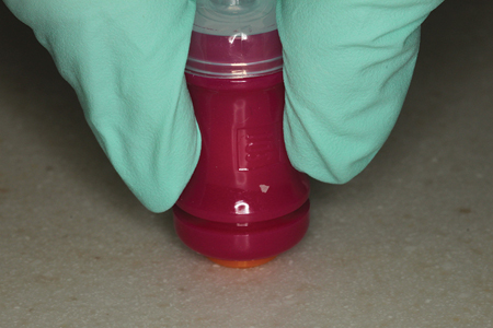
Fig. 1 A-J
An erupting mandibular first molar is sealed with Fuji TRIAGE (white) (GC America)


Fig. 2 A-G
An erupting maxillary first molar is sealed with Riva Protect (pink) (SDI Australia)


Conclusion
Fissure sealant application is an excellent proactive dental treatment. It is an underused treatment because of the difficulties in isolation with resin sealants and the unwelcome surprises of advanced decay that is sometimes found under failed resin sealants. Glass ionomer sealants offer the advantages of easier isolation and the ionic exchange of fluoride and other minerals to help in the mineralization of the immature tooth surface. It is time to bring fissure sealants back out, as proactive intervention treatments for our young patients, this time with patient friendly glass ionomer materials. OH
Oral Health welcomes this original article.
References
- Muthu MS, Sivakumar N. Pediatric dentistry. Principles and Practice. 1st ed. New Delhi: Elsevier; 2009
- Gore DR. The use of dental sealants in adults: a long-neglected preventive measure. Int J Dent Hyg 2010; 8: 198-203
- Cueto El, Buonocore MG. Sealing of pits and fissures with an adhesive resin: its use in caries prevention. J Am Dent Assoc 1967; 75(1):121-8
- Ekstrand KR, Christiansen J, Christiansen ME. Time and duration of eruption of first and second permanent molars: a longitudinal investigation. Community Dent Oral Epidemiol. 2003 Oct; 31(5):344-50
- Antonson DE. Imagine a world without occlusal caries: are glass ionomer sealants the answer? Oral Health Journal 2012 Dec; 31-36
- Berg JH. Glass ionomer cements. Pediatric Dent 2002; 24(5):430-438
- Ahovuo-Saloranta A, Hiri A, Nordblad A, Makela M, Worthington HV. Pit and fissure sealants for preventing dental decay in the permanent teeth of children and adolescents. Cochrane Database Syst Rev 2008; 4:CD001830
- Bishara SE, Oosombat C, Ajlouni R, Denehy G. The effect of saliva contamination on shear bond strength of orthodontic brackets when using a self-etch primer. Angle Orthod 2002; 72:554-557
- Smith DC. Development of glass-ionomer cement systems. Biomaterials 1998; 19:467-478
- Yengopal V, Mickenautsch S, Benzerra AC, Leal SC. Caries-preventive effect of glass ionomer and resin-based fissure sealants on permanent teeth – a meta analysis. J Oral Sci 2009; 51:373-382
- Mickenautsch S, Yengopal V. Caries-preventive effect of glass ionomer and resin-based fissure sealants on permanent teeth: an update of systematic review evidence. BMC Research Notes 2011; 4:22
- Mickenautsch S, Yengopal V. Retention loss of resin-based fissure sealants – a valid predictor for clinical outcome? The Open Dentistry Journal 2013; 7:102-108
- Niederman R. Glass ionomer and resin-based fissure sealants: equally effective? Evid Based Dent 2010; 11(1):10
- Edwina A. M. Kidd. Essentials of Dental Caries: The Disease and its Management. Oxford University Press p 170, June 30, 2005
- Edwina A. M. Kidd. Dental Caries: The Disease and its Clinical Management. John Wiley & Sons. P299, Apr 11, 2008
- Sachin S. Glass ionomer cement and resin-based fissure sealants are equally effective in caries prevention: a critical summary of Yengopal V, Mickenautsch S, Benzerra AC, Leal SC. Caries-preventive effect of glass ionomer and resin-based fissure sealants on permanent teeth – a meta analysis. J Oral Sci 2009; 51:373-382. JADA 2011 May; 142(5):551-552
- Antonson, SA, Kilinc E, Antonson, DE. Depth of Penetration of Fissure Sealants on Contaminated Enamel Surface. J Dent Res 2006; 85(Spec Iss B):1580
About the Authors

Dr. Fay Goldstep has lectured nationally and internationally on Proactive/Minimal Intervention Dentistry, Soft-Tissue Lasers, Electronic Caries Detection, Healing Dentistry and Innovations in Hygiene. She has been a contributing author to four textbooks and has published more than 100 articles. She sits on the editorial board of Oral Health. Dentistry Today has listed her as one of the leaders in continuing education since 2002. Dr. Goldstep is a consultant to a number of dental companies, and maintains a private practice in Richmond Hill, Ontario. She can be reached at goldstep@epdot.com.
 Cathy Delios graduated from Georgian College in 2000 and has been in private practice for the last 18 years. She is very interested in nutrition and overall health and wellness, which provides endless learning opportunities. Cathy is an avid reader and sports fan and loves to exercise when she is not busy with her husband and two active sons.
RELATED ARTICLE: Imagine a World Without Occlusal Caries: Are Glass Ionomer Sealants the Answer?


