Over the past four decades, millions of all-ceramic restorations, both veneers and crowns, have been cemented (or bonded) in place in order to satisfy the ever-growing demand for aesthetic smiles. Dentists have invested many hours of continuing education to enhance their diagnostic and clinical capabilities, while researchers and dental companies have made sustained efforts to improve the mechanical, chemical, and optical properties of the materials used in these highly demanding cases. Most of these efforts correspond to first-time treatments, however, where the operating conditions differ greatly from situations where old restorations need to be replaced.
Ceramic restorations can fail for a number of reasons, including ceramic fracture, defective margins, secondary caries, discoloration, receding gingiva, periodontal compromise and/or overall patient dissatisfaction with the esthetic appearance.1
Guidelines for removing existing ceramic restorations are scarce. PFM crowns can be carefully removed by cutting guide grooves and then manipulating the underlying metal with crown-spreading pliers or crown-dilating forceps. Ceramic restorations which have been adhesively cemented should NOT be cut open and similarly manipulated due to the high risk of fracturing the bonded tooth structure underneath.2 It is safer to remove existing ceramic restorations with a high-speed handpiece with a diamond bur. The major problem with this technique lies in the color-blending properties of both the ceramic and the resin cement, which make them very difficult to differentiate from the underlying dental tissues, particularly under the wet conditions required for high-speed removal. The procedure, thus, becomes both stressful and time-consuming for the dentist, while the risk of causing iatrogenic damage to the underlying tooth structure remains.2
The ideal ceramic removal method debonds old restorations, hopefully intact, quickly and with no damage to the tooth structures.3 In fact, this method has existed for years in the form of laser technology. However, despite the rapid popularization of these procedures in medicine, laser use is still not common practice in the dental field.
Laser Removal of Ceramic Restorations
Several types of lasers can be used for a wide range of more than 40 applications in dentistry. Diode lasers, for example, are most often used on soft tissues for procedures such as gingival contouring, frenectomy, implant exposure, removal of hyperplastic tissues, etc. The erbium-doped:yttrium aluminum garnet (Er:YAG) laser, on the other hand, is characterized by having applications on both soft and hard tissues, making it suitable for caries removal, enamel etching, osteotomy, root resection, etc. Due to the Er:YAG wavelength’s capacity to be absorbed by materials that contain water or residual monomers, it can also be used to degrade resinous materials, such as resin cements, thus making it ideal for removing veneers and all-ceramic crowns.3,4
Two mechanisms enable this Er:YAG laser activity: First is Thermal Ablation, which is the fast vaporization of the resinous adhesive caused by the laser energy. Second is Photoablation, which is the laser energy’s interaction with the resinous adhesive, that increases the resin molecules’ energy levels up to the level of their dissociation, resulting in decomposition of the material.5
The debonding process is affected by the laser parameters (power, pulse duration, frequency, and lasing time), as well with other factors related to the ceramic material such as chemical composition, shade, opacity, and thickness. For example, thinner restorations are removed more quickly than thicker ones. In any case, most restorations can be removed with just a few minutes of laser exposure. It is important to remember, however, that there will be a temperature increase during the debonding process. Therefore, using the recommended laser parameters and sufficient air-water cooling are fundamental to avoid causing pulpal damage.6,7
In terms of ease of use, current laser equipment is more user-friendly than ever. As with any other new technology, there is a learning curve but it is definitely worth the effort. The following case reports will demonstrate the application of an Er:YAG laser to removing ceramic restorations in the esthetic zone.
Case 1
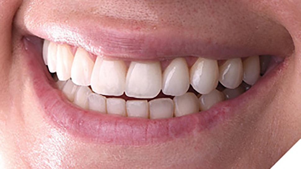
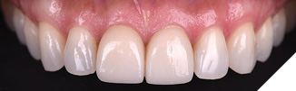
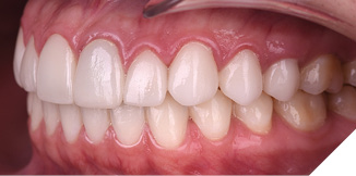
The 45-year-old female patient, presented with a complaint concerning the esthetic appearance of two upper central veneers which had been placed one year prior. Upon clinical examination, she had good oral health and a stable occlusion. It was noted that the two ceramic veneers were indeed unesthetic in color, translucency, size, and shape (Figs. 1,2). After radiographic and photographic assessment, the proposed treatment plan consisted of debonding the ceramic veneers using an Er:YAG laser (LiteTouch, Light Instruments, Yokneam, Israel) and replacing them with new lithium disilicate IPS e.max veneers (Ivoclar Vivadent, Mississauga ON).
Fig. 1
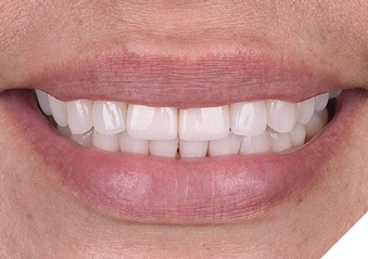
Fig. 2
Despite tooth vitality, no anesthesia was required for the debonding procedure. The Er:YAG laser was set to the following parameters: 20 Hz, 250 mJ, and 5 W, with air/water spray irrigation. The laser tip was placed perpendicular to the tooth surface at a distance of 5mm and activated, making circular motions along the surface of the veneer for a period of 2 minutes. Both veneers were finally removed using a lever movement with a spatula at the cervical junction. The tooth preparations were minimally adjusted with fine diamond burs and round discs, and polyvinyl siloxane dental impressions were taken using #00 retraction cord.
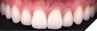
In the following appointment, the new veneers were tried in and after patient approval they were cemented into place using customary bonding protocol (Figs. 3,4). The clinical video is available here.
Fig. 3
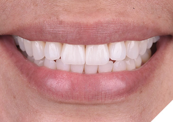
Fig. 4
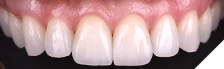
Case 2
A 31-year-old female patient presented with gingival inflammation and pain related to ceramic veneers and a crown placed 6 months earlier (Figs. 5,6). The radiographic assessment revealed a failing endodontic treatment on the left central incisor. To reduce inflammation of the surrounding gingiva, the ceramics were recontoured at the gingival margins using fine burs and polishing points. After two weeks, periodontal health had improved considerably and the Er:YAG laser was used with the same protocol as described for Case 1 to remove the existing restorations.
Fig. 5A
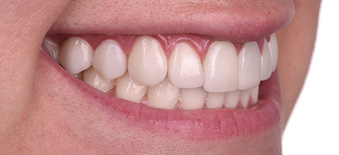
Fig. 5B
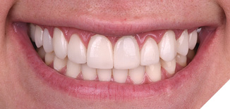
Fig. 5C
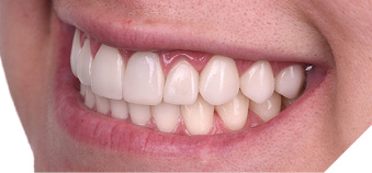
Fig. 6A
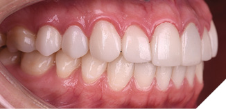
Fig. 6B
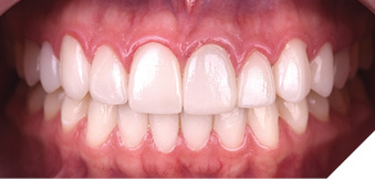
Fig. 6C
However, in this case, the veneers did not pop out intact but disintegrated during the removal process. This may be due to the thickness or the composition of the material (seemingly felspathic porcelain). The clinical video is available here.
As expected, the remaining tooth structure was entirely intact and a new set of 9 veneers and a crown were fabricated. Two weeks after cementation, an apicectomy was performed on the left central incisor using the same laser.
At the two-month post-op control, soft tissues were healthy, and the patient indicated her complete satisfaction with the results (Figs. 7,8,9).
Fig. 7A
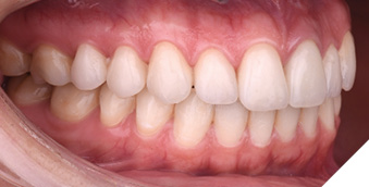
Fig. 7B
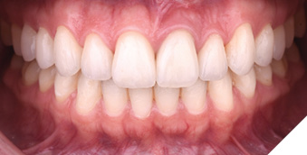
Fig. 7C
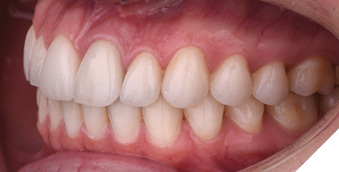
Fig. 8A
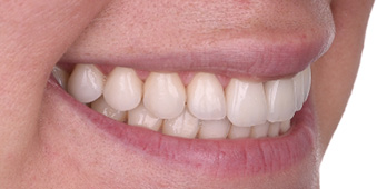
Fig. 8B
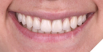
Fig. 8C
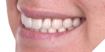
Fig. 8D
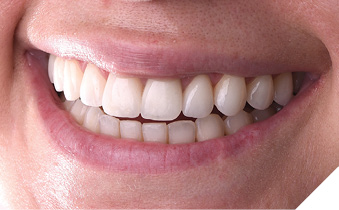
Fig. 9A
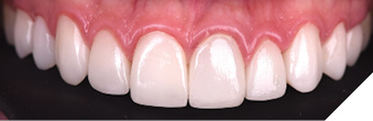
Fig. 9B
Conclusion
Dental practitioners often need to remove old ceramic restorations safely and conveniently. Unfortunately, the application of laser technology for this purpose is still not common practice. Laser removal of ceramic restorations is easy, safe, preserves tooth structures, generates a positive patient experience, and saves chairside time. These are all substantial clinical benefits.
Additionally, the removal of cemented ceramics is but one of more than twenty applications of the same laser equipment, making it a worthy investment. Finally, any technology capable of reducing the stress levels to which dentists are exposed every day should be more valued. 
Oral Health welcomes this original article.
References
- Alenezi A, Alsweed M, Alsidrani S, Chrcanovic BR. Long-Term Survival and Complication Rates of Porcelain Laminate Veneers in Clinical Studies: A Systematic Review. J Clin Med. 2021 Mar 5;10(5):1074
- Spath A, Smith C. Removal of Modern Ceramics. Compend Contin Educ Dent. 2017 May;38(5):326-333. PMID: 28459251
- Buu N, et al. Er:YAG laser debonding of porcelain veneers. Laser in Dentistry. 2010;7549:754909-754912.
- Morford CK, Buu NC, Rechmann BM, Finzen FC, Sharma AB, Rechmann P. Er:YAG laser debonding of porcelain veneers. Lasers Surg Med. 2011 Dec;43(10):965-74
- El-Damanhoury HM, Salman B, Kheder W, Benzina D. Er:YAG laser debonding of lithium disilicate laminate veneers: effect of laser power settings and veneer thickness on the debonding time and pulpal temperature. J Lasers Med Sci. 2022;13:e57
- Ghazanfari R, Azimi N, Nokhbatolfoghahaei H, Alikhasi M. Laser aided ceramic restoration removal: a comprehensive review. J Lasers Med Sci. 2019;10(2):86-91
- Sari T, Tuncel I, Usumez A, Gutknecht N (2014) Transmission of Er: YAG laser through different dental ceramics. Photomed Laser Surg 32:37–41
About the Author

Dr. Paola Ochoa graduated with top honors from Cayetano Heredia University and Universidad Científica del Sur (Occlusion and Oral Rehabilitation). Following extensive training in Brazil and the United States, she co-founded Infinity Dental Clinic and Infinity Advanced Dental Learning in Lima, Peru. Dr. Ochoa is a Fellow of the American Society for Dental Aesthetics and a Diplomate in the American Board of Aesthetic Dentistry, and employs an interdisciplinary, facially driven, and minimally invasive philosophy, utilizing digital tools for planning and communication.