
Introduction
Imagine the following scenario. Due to the COVID-19 pandemic, you can only treat patients with acute emergencies, in order to help slow or stop the spread of the coronavirus. However, a patient calls you complaining of a severe tooth ache or swelling that you must somehow manage. You may wonder if you could or should manage this with over the counter or prescription medications, or if you have to treat this invasively and immediately. In many cases, a true acute emergency of endodontic origin needs to be clinically managed right away.1
This article will review how you should manage an emergency of endodontic origin and how to do so efficiently. I will divide this into the following key steps: diagnosis, anesthesia (especially of “hot teeth”), canal instrumentation and post-op pain and infection management. All while keeping in mind COVID-19 considerations.
Diagnosis and COVID-19 Considerations
The first and obvious step involves figuring out what is and where is the source of the pain, and if it is tooth (endo) related. Make sure to rule out non-endo related symptoms (ie periapical inflammation due to parafunction or pain of periodontal origin). If possible, diagnose as much as possible by phone, in order to minimize direct patient interaction. If needed, perform all the routine diagnostic endodontic tests (ie pulp tests, percussion, palpation, biting on a tooth slooth) clinically. If the emergency is found to be of endodontic origin and you’ve localized the problem tooth, and this cannot be managed by medications, then proceed with root canal treatment.1
If indeed this patient needs to be managed immediately, then make sure to follow your local regulatory and public health bodies’ guidelines for treatment, with respect to COVID-19. It is important to ensure that you and your staff have the proper personal protective equipment and it is also important to minimize or eliminate the production of contaminated aerosols during treatment. Fortunately, in endodontics this is relatively easy to achieve by isolating the tooth, post-anaesthesia and pre-access, with rubber dam, and then rinsing the crown with chlorhexidine or hypochlorite.2
Anesthesia
This is often the most difficult and important aspect of the emergency. Without profound pulpal anesthesia, you’ll have a hard time proceeding. Insufficient or lack of anesthesia may turn the appointment into a memorable nightmare for the patient and be very stressful for everyone.
Maxillary Teeth
Traditional local infiltration will suffice for most cases in the maxilla. But, make sure to have the needle tip deposit the anesthetic superior to the apices of the involved tooth. For instance, if you are treating a maxillary first molar, try not to hit the zygoma with the needle. This may result in deposit of the anesthetic inferior to the site of innervation to the involved tooth and compromise pulpal anesthesia.
“Hot Teeth”
Arguably, the most difficult tooth to anaesthetize is the infamous “hot tooth” and are well known to remain “alive” even after numerous carpules of local anaesthetic.
It has been shown by numerous clinical studies that mandibular blocks will only provide pulpal anesthesia in about HALF the cases with irreversible pulpitis. So, if your patient presents with acute lingering pain to cold, there’s only about a 50% chance that any mandibular block will suffice.3 This means that in many cases, you will require supplemental anesthesia. (See Table 1). These include the following:
Table 1
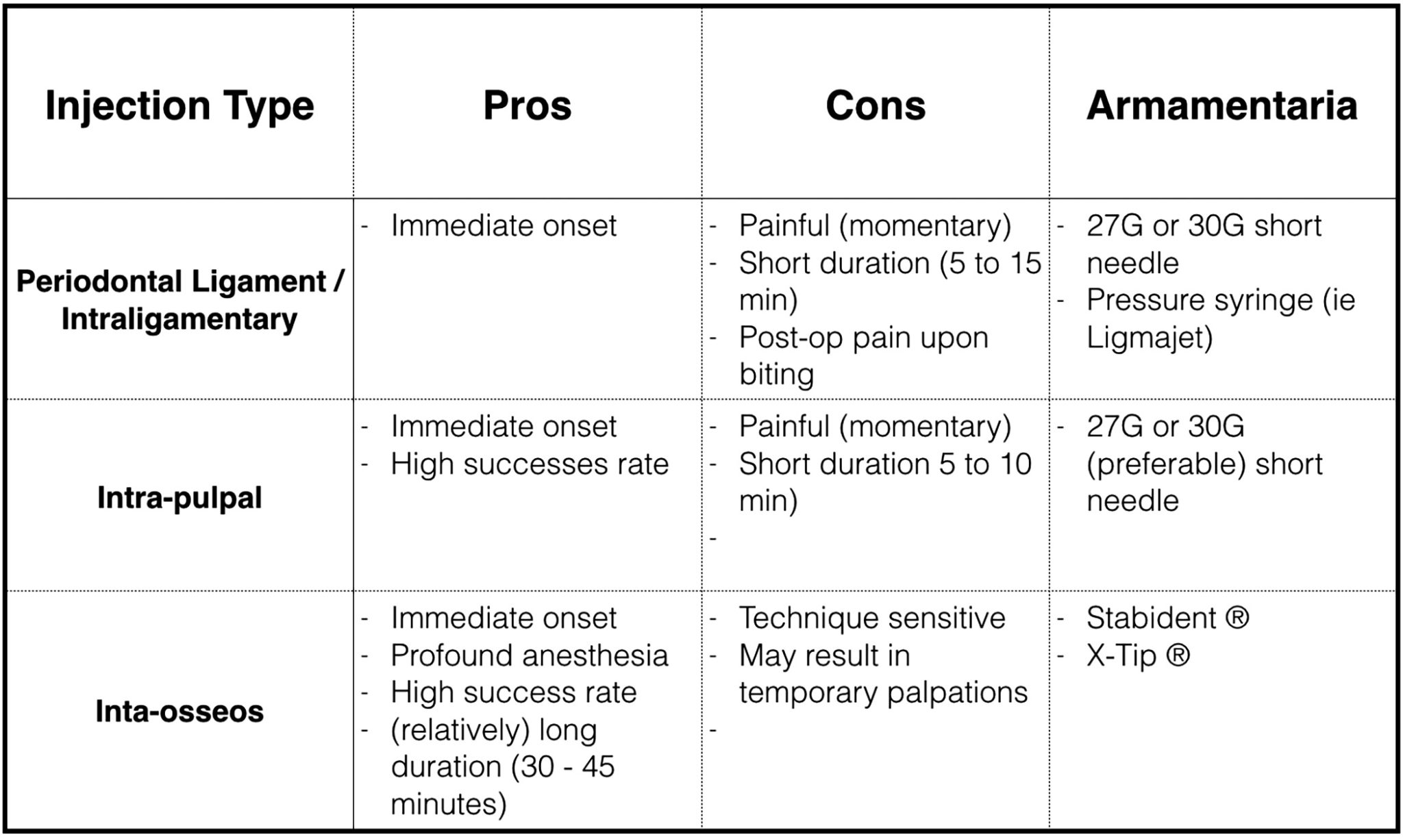
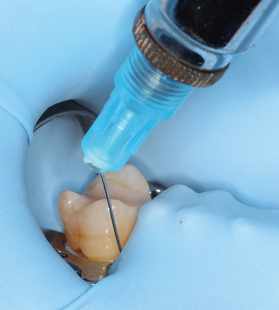
Periodontal Ligament Injection (PDL), AKA Intraligamentary Injection
This is performed using a 30 gauge (or possibly 27G) short needle that is placed at the mesial or distal line angles of the tooth’s periodontal crevice and with the bevel towards the tooth. This could be performed with rubber dam isolation. (Figs. 1 & 2). Under pressure, a small amount of anesthetic is injected. Onset is quick but the anesthesia is relatively short lasting. This injection is often uncomfortable and patients may complain of the tooth feeling elevated, post-op. PDL injections may be used as supplemental anesthesia for maxillary and mandibular teeth.4,5
Fig. 1

Fig. 2
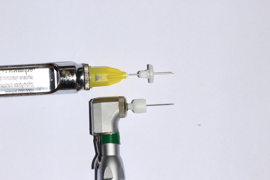
Intra-Osseous
This type of injection has been shown to provide exceptionally high rates of immediate and profound pulpal anesthesia, including in “hot teeth”. It is used primarily for mandibular posteriors. It requires a pilot hole to be drilled with a special slow speed instrument (perforator) into the cortical bone between the teeth at the base of the papilla and angled apically. Into the pilot hole, a corresponding short needle is inserted and the anesthetic is slowly deposited. (Figs. 3, 4 & 5). Its duration is approximately 30 to 45 minutes. Also, it is recommended that an anesthetic with little epinephrine (ie 1:200,000 epi) be used to minimize heart palpitations.4 The two options available are the “X-Tip®” (Dentsply Sirona) and “Stabident®” (Fairfax Dental, Miami). (Figs. 6 & 7).
Fig. 3
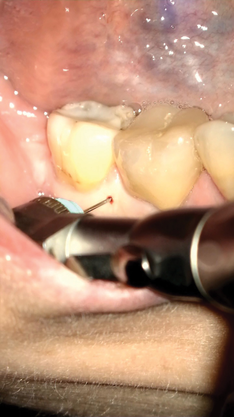
Fig. 4

Fig. 5

Fig. 6
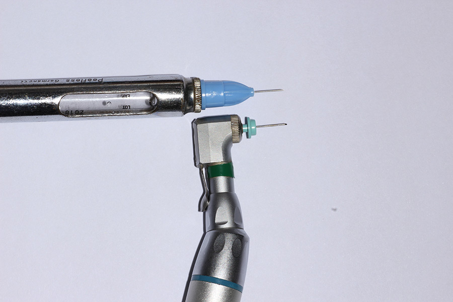
Fig. 7
At this point, I suggest pulp testing the tooth prior to your access, to make sure it is anaesthetized. There is nothing worse than a nervous emergency patient with a hot tooth jumping from pain midtreatment. And, once anesthetized, and as stressed earlier, you must isolate the tooth with rubber dam prior to initiating the root canal access.
Intra-Pulpal
This is often considered the anesthetic of last resort. When performed properly, it is immediate but will only provide about 10 minutes of profound anesthesia. As with PDL injections, it is very painful when administered. (6) The needle should engage in the canal and for this to work, you must obtain resistance when injecting the anesthetic. (Figs. 8 & 9).
Fig. 8
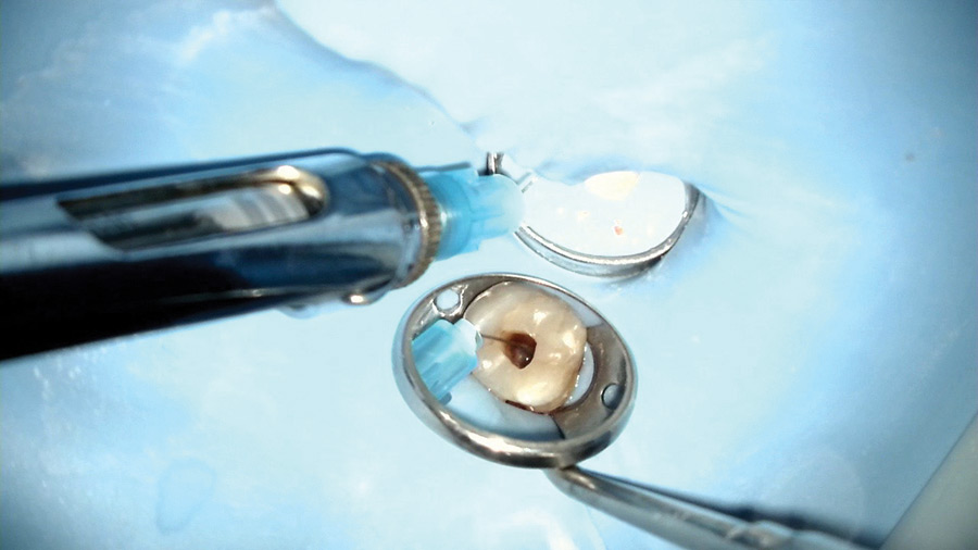
Fig. 9

Soft Tissue Abscess of Endodontic Origin
If your patient presents with a soft tissue swelling that is fluctuant, you should perform an incision and drainage (“I & D”). (Figs. 10-14). You should use a sharp scalpel and penetrate the “pointing” part of the abscess until you hit bone. A curved hemostat should be used to expand the incision site and help provide a way for the purulence to drain. For significant swellings, a drain should be placed. This could be accomplished with a clean rubber dam cut in the form of the letter “T” with the top of the letter inserted into the incision site and sutured into place.
Fig. 10

Fig. 11
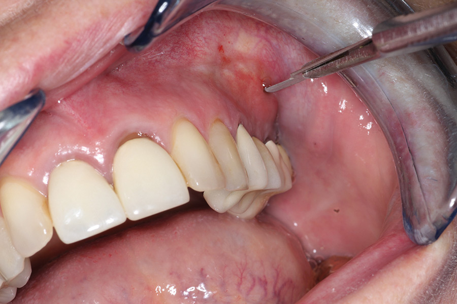
Fig. 12
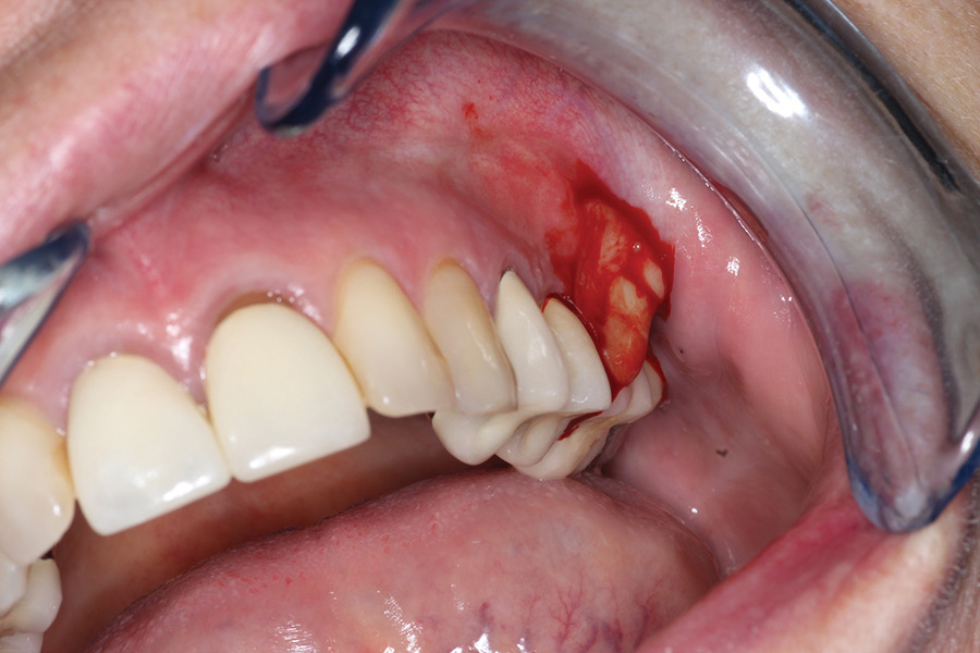
Fig. 13

Fig. 14

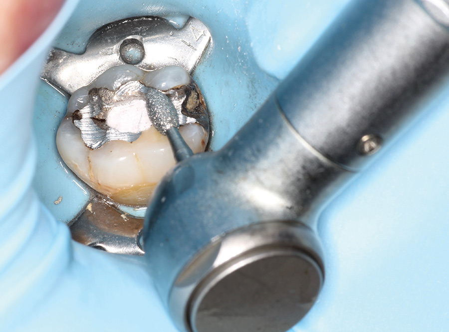
Root Canal Instrumentation
By now you have diagnosed and identified the endo problem, anesthetized the patient and accessed the tooth. Next comes the canal instrumentation. It may be difficult, in the heat of the moment, but do make sure to locate and treat all the canals. This is much easier if magnification with enhanced illumination is incorporated. Dental loupes with a headlight or a dental microscope are exceptionally helpful, if not priceless.
If endodontic treatment is not to be completed that day, it is very important to completely instrument each canal. Anything less means leaving behind inflamed or infected pulp tissues. A pulpectomy by definition involves complete pulp extirpation and canal instrumentation.1 Furthermore, the canals should be medicated with calcium hydroxide between appointments.
As you are in the midst of managing an emergency, it is extremely helpful to use an instrumentation system that is simple, efficient and easy to use. The trend in endodontic instrumentation has been to achieve the same or better clinical and biological outcomes, with fewer steps and instruments. Single file mechanized NiTi instrumentation is a relatively recent addition to endodontics. Thanks to its simplicity and efficiency, it is well suited for, amongst other things, emergency root canal treatments. An example is the WaveOne Gold reciprocating NiTi file. (Dentsply Sirona, Tulsa, OK) This file system has been shown to be safer, simpler and more efficient to use than many rotary NiTi systems.8 (Fig. 17). With respect to rotary NiTi files, there are newer systems that still require multiple files, but which work more efficiently and require slightly fewer NiTi files than traditional systems such as the ESX series of files (Brasseler USA Dental, Savannah, GA).
Fig. 15
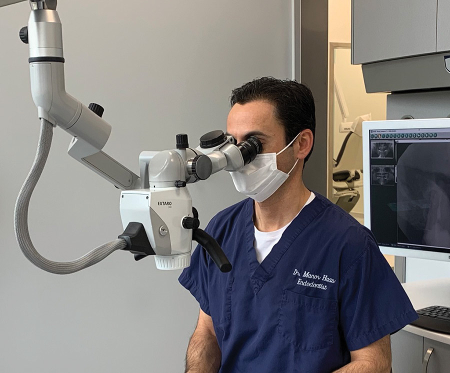
Fig. 16
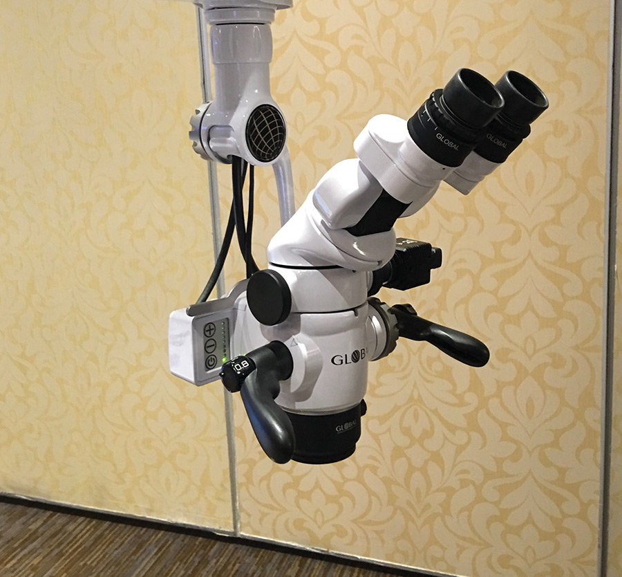
Fig. 17

Post-Op Management
Occlusion
From a pain management perspective and if possible, it is recommended to reduce the occlusion of the involved tooth. The intention is to prevent post-op occlusal trauma to the tooth and periapical area that needs to be left alone to settle, so-to-speak. This is especially important in patients with parafunction habits who may be more likely to traumatize the problem tooth. (Fig. 18). In these times of COVID-19, you might not know when you’ll be able to see your patient again. Hence, I would also recommend you temporize the access with a permanent restoration.
Fig. 18
Pain Management
As a rule of thumb, if a patient presents with pre-op pain (except for pain to cold and hot), they are likely to experience post-op pain.7 In turn, you should prepare your patient to this and also provide them with analgesics suitable for their level of pain. For a healthy patient, these may range from 600mg of Ibuprofen every 6 hours for mild pain, to 1 to 2 Tylenol 3 every 4 to 6 hours, for more severe pain.
Infection Management:
It is rare that post-op anibiotics will be indicated for endodontic emergencies, so long as the etiology is managed endodontically. That is, unless the patient presents with a space infection or systemic symptoms.9 For routine cases and patients, the antibiotic of choice is Amoxicillin (500mg, q8h). For patients with Penicillin allergies, Clindamycin is recommended (300mg, q6h). Both for a week.
Conclusion
One of the greatest challenges in clinical dentistry is managing endodontic emergencies. The coronavirus has only complicated this. So, when treatment is performed, it should be done efficiently and under profound pulpal anesthesia and with reduction or elimination of contaminated aerosols by using rubber dam isolation. And all this should be done while using the recommended personal protective equipment.
Oral Health welcomes this original article.
Disclosure: Dr. Haas reports no disclosures.
References
- Hargreaves KM, Berman LH. Cohen’s Pathways of the Pulp. 11th ed. St. Louis, MO: Elsevier;706, 2016.
- Meng L. et al, Coronavirus Disease 2019 (COVID-19): Emerging and Future Challenges for Dental and Oral Medicine. J Dent Res 00(0), 2020.
- Claffey E, Reader A, et al. Anesthetic efficacy of articaine for inferior alveolar nerve blocks in patients with irreversible pulpitis. J Endod 30 (8), 568, 2004
- Malamed SF. Supplemental injection techniques. Handbook of Local Anesthesia. 5th ed. St. Louis: Mosby; 2004.
- Moore PA, et al.: Periodontal ligament and intraosseous anesthetic injection techniques, J Am Dent
Assoc 142, 2011. - Hargreaves KM, Berman LH. Cohen’s Pathways of the Pulp. 11th ed. St. Louis, MO: Elsevier;706, 2016.
- Hargreaves KM, Keiser K: New advances in the management of endodontic pain emergencies. J Calif Dent Assoc 32:409, 2004.
- Abn,S, Kim H-C, Kim E, Kinematic Effects of Nickel-Titanium instruments with reciprocating or continuous rotation motion: a systematic review of In Vitro studies. J Endod 42:1009, 2016.
- Henry M, Reader A, Beck M: Effect of penicillin on postoperative endodontic pain and swelling in symptomatic necrotic teeth. J Endod 27:117, 2001.
About the Author
 Dr. Haas is a certified specialist in endodontics and lectures internationally. He is a Fellow of the Royal College of Dentists of Canada and is on staff at the University of Toronto Faculty of Dentistry and the Hospital for Sick Children. He maintains a full-time private practice limited to endodontics and microsurgery in Toronto. Dr. Haas is a regular contributor to dental journals and online forums. He can be reached via the website: www.HaasEndoEducation.com
To view more COVID-19 news as it pertains to the dental profession, please click here.


