Abstract
Arteriovenous malformations (AVMs) are rare vascular lesions composed of arteries and veins with congenital structural defects. Their ambiguous clinical and radiographic presentations make their recognition and diagnosis challenging for dental professionals. A healthy 14-year-old male presented to The Hospital for Sick Children (SickKids) in Toronto, Ontario with a chief concern of hematogenous malignancy that had been diagnosed by a community oral radiologist. The dental professionals and family doctor first involved in the case requested the family present to the SickKids Emergency Department to access hospital resources more quickly. While receiving hospital care and awaiting his next consult, a near catastrophic hemorrhagic event occurred. Fortunately, the patient was in the SickKids Emergency Department when this event took place. Had this event happened in any other setting, the outcomes may have been fatal. It is therefore crucial for dental professionals to recognize these lesions, and appropriately refer patients with AVMs to the appropriate healthcare provider. It is also important for one to be familiar and comfortable with emergency management of such hemorrhagic events.
Introduction
Vascular malformations constitute a group of congenital conditions characterized by an inappropriate development of vessels (Kumar et al.). These disorders can be subdivided into four main categories, each one differentiated by the type of vessels affected by the abnormality. We therefore recognize capillary malformations, venous malformations, lymphatic malformations and arteriovenous malformations (“Vascular Lesions | SSM Health”). For the purposes of this case report, arteriovenous malformations will be discussed in more detail.
Arteriovenous malformations (AVMs) are vascular lesions composed of arteries and veins with congenital structural defects. The vessels at the origin of an arteriovenous malformation have endothelial cells with normal turnover rates (Kumar et al.). It is known that AVMs may increase in size dramatically during times of puberty, pregnancy or after a trauma. It is also suspected that hormonal signaling pathways may be involved in collateral proliferations, however more research is necessary to understand these particular mechanisms (Bhandary et al.).
AVMs may occur anywhere in the body. A recent study by Su et al. states that although AVMs represent 1.5 per cent of all vascular anomalies, 50 per cent of those lesions occur in the oral and maxillofacial region (Cjdr.Quintessenz.De, 2019, https://cjdr.quintessenz.de/cjdr_2014_02_s0085.pdf. Accessed 17 Oct 2019).
Due to their ambiguous and often varied presentations, AVMs can be easily misdiagnosed and consequently improperly managed. Given the potentially fatal outcomes that can accompany lesions of this nature, it is fundamental that oral healthcare professionals are competent in identifying, managing and/or referring patients with suspected AVMs.
Case Report
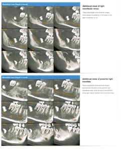
An otherwise healthy 14-year-old male presented to the Emergency Department at The Hospital for Sick Children (SickKids) in Toronto, Ontario with the chief concern of dental pain localized to the right posterior mandible. The patient had a one-year history of progressive facial swelling (Figs. 1-4) with right sided lower lip and chin hypoesthesia that had developed approximately one month prior to this hospital presentation. At the time, the patient denied a history of fevers, chills, night sweats or recent weight loss. He had previously visited his community dentist for a routine dental exam and cleaning when it was noted that tooth 47 was mobile. The dentist proceeded to take a panoramic radiograph which revealed an apically displaced, unerupted tooth 48 in the posterior right mandible with a large radiolucent lesion spanning from the posterior mandible to the distal root of tooth 47 (Fig. 5). The patient was then referred to a community orthodontist who subsequently referred the patient to an oral radiologist for diagnostic imaging. A CBCT scan was completed and a report was given to the referring orthodontist, who then delivered the findings to the patient and his family (Figs. 6 & 7). In the radiology report, the diagnosis given was a hematogenous malignancy, and the differential diagnoses included lymphoma leukemia, plasmocytoma and Langerhans cell histiocytosis.
Fig. 1
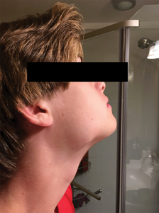
Fig. 2
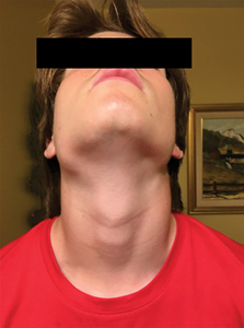
Fig. 3
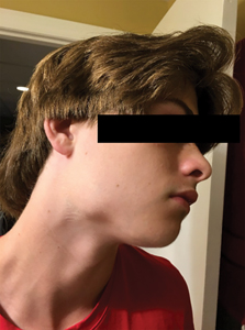
Fig. 4

Fig. 5
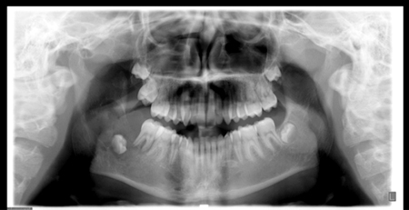
Fig. 6
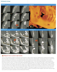
Fig. 7
The dental professionals and family doctor involved in the case requested the family present to the Emergency Department at SickKids to facilitate quicker access to hospital resources, including diagnostic imaging and blood tests. After presenting to the Emergency Department, a proper workup was ordered. A consult with the Oral and Maxillofacial Surgery (OMFS) Service at the Department of Dentistry was booked for the team’s next clinic day for further investigation and possible biopsy if deemed appropriate. At the time of the patient’s consult with the OMFS team, it was decided not to biopsy the lesion due to the possibility of the lesion being vascular in nature, which would result in an unpredictable bleeding event. The patient was referred to Plastic Surgery for further management and definitive treatment of the patient’s mandibular lesion. The patient and his family were advised to present again to the SickKids Emergency Department should the patient detect any bleeding in the mouth, increase in size or increase in jaw pain while waiting for their consultation appointment with Plastic Surgery. The patient and his mother presented for a second time to the Emergency Department, after detecting some blood while brushing the teeth one night before bed. During this visit, the patient sneezed while waiting to be seen by an emergency department team member, shortly after being triaged. At this time, the patient began to haemorrhage from the mouth. It was estimated that the patient lost approximately one litre of blood in a 45-minute period, during which time blood transfusions were delivered and an emergency operating room was booked. The patient was brought to a SickKids operating room where the perforated vascular lesion was glued and embolized by medical personale. Since this time, the patient has been closely followed and managed by the Plastic Surgery team at SickKids, where the definitive treatment originally included mandibular resection and reconstruction using free fibula.
After having a successful follow up appointment two months later, it was noted that the patient did well with embolization, and since there is increasing data that embolization may be sufficient as definitive treatment for young patients with AVMs, the free fibula reconstruction has been deferred for now. Embolization as a definitive conservative treatment has been shown to have a 70 percent success rate in the literature for AVMs developing before adolescence (R. Spreafico).
It is important to note that reconstructive jaw procedures completed at SickKids are primarily managed by the Plastic Surgery service and not Oral Maxillofacial Surgery. This situation is unique to SickKids and is the reason the Oral Maxillofacial Surgery team referred the patient to this particular hospital service.
Discussion
Vascular lesions often present as ambiguous entities. It is therefore imperative to know the signs and symptoms to ensure proper referral and subsequent diagnosis and management. Characteristics of vascular lesions include pain (51%), functional compromise (27%), swelling (24%) and disfigurement (21%) (Mathes et al.). Additionally, the majority of patients with a vascular anomaly present with a colour change, where the lesion will have a characteristic reddish-purple appearance that will be distinct from the surrounding pink alveolar mucosa and gingiva (“Vascular Lesions | SSM Health”). Since the lesion is vascular, if the mass is large enough, it may also pulsate.
If you suspect a patient of having an oral AVM, you should refer the patient to a hospital based oral maxillofacial surgeon for further investigation and proper management. Ultrasound investigations, CT and MRI scans, as well as blood tests should be completed to assist with proper diagnosis. Signs and symptoms of a bleeding event should be discussed at length with the patient and their family so they know when it is appropriate to present to an emergency department. It must be stressed that even if a small amount of bleeding is detected during toothbrushing in the area of the suspected or confirmed vascular oral lesion, presentation to an emergency department is indicated, as was the case in the scenario described here.
In the event a patient with a suspected or confirmed AVM experiences a bleeding episode in your dental office or somewhere else outside of a hospital setting, ensuring the airway is maintained is paramount. Calling 911 and maintaining direct pressure against the site of perforation with gauze should be completed after the airway is secured and maintained until emergency services arrive.
Conclusion
In conclusion, AVMs in the oral cavity are rare and can have life threatening consequences. It is crucial to properly recognize and refer patients with suspected AVMs to the appropriate healthcare providers to ensure appropriate investigation, diagnosis and management in a timely fashion.
Oral Health welcomes this original article.
References
- Kumar, Ashok et al. “Arteriovenous Malformation Of Face”. Contemporary Clinical Dentistry, vol 8, no. 3, 2017, p. 482. Medknow, doi:10.4103/ccd.ccd_100_17.
- “Vascular Lesions | SSM Health”. Ssmhealth.Com, 2019, https://www.ssmhealth.com/cardinal-glennon/pediatric-plastic-reconstructive-surgery/hemangiomas.
- Cjdr.Quintessenz.De, 2019, https://cjdr.quintessenz.de/cjdr_2014_02_s0085.pdf. Accessed 17 Oct 2019.
- Bhandary, B Satheesh Kumar et al. “Traumatic Arteriovenous Malformation Of Cheek: A Case Report And Review Of Literature”. An International Journal Of Otorhinolaryngology Clinics, vol 5, no. 3, 2013, pp. 173-177. Jaypee Brothers Medical Publishing, doi:10.5005/jp-journals-10003-1138.
- Mathes, Erin F. D. et al. “Clinical Characteristics And Management Of Vascular Anomalies”. Archives Of Dermatology, vol 140, no. 8, 2004. American Medical Association (AMA), doi:10.1001/archderm.140.8.979.
- R. Spreafico, F. Parmigiani. “Arterio-Venous Malformation Of The Mandible. Case Report And Review Of Literature”. Pubmed Central (PMC), 2019, https://www.ncbi.nlm.nih.gov/pmc/articles/PMC5066472/. Accessed 22 Oct.
About the Author
 Dr. Mallory Laframboise is a graduate of McGill University Faculty of Dentistry, and a current dental resident in the Department of Dentistry at the Hospital for Sick Children in Toronto, Ontario, Canada.
 Dr. Audrey Lacombe is a graduate of La Faculté de Médecine Dentaire de l’Université de Montréal, and a current dental resident in the Department of Dentistry at the Hospital for Sick Children in Toronto, Ontario, Canada.
RELATED ARTICLE: Arteriovenous Malformation of the Left Maxillary Sinus: Case Report