Note from editor
Oral Health is proud to introduce a brand-new series by Dr. Bruce Pynn and Dr. Hagen Klieb, featuring a curated collection of rare and compelling cases. This series aims to raise awareness and deepen understanding of unique and unusual pathologies within the dental profession.
How would you diagnose this case?
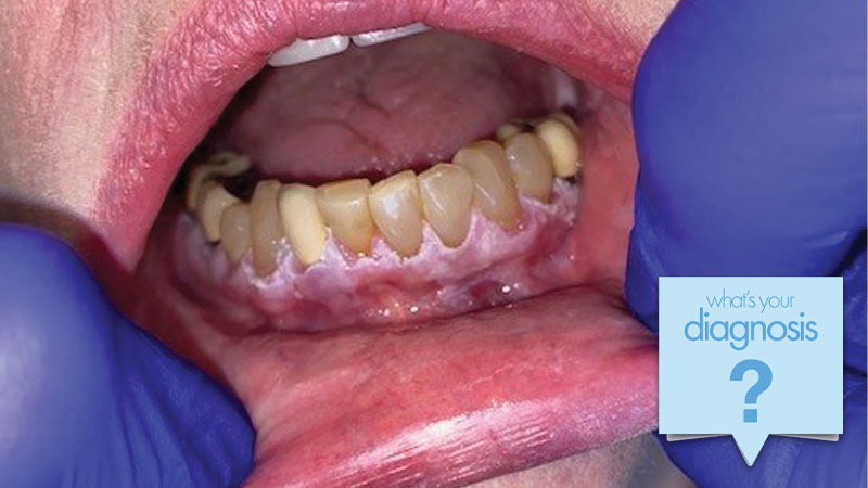
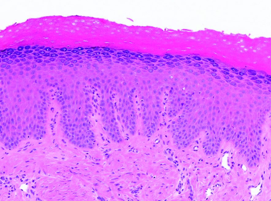
A 68-year-old woman was referred to the oral pathology service for assessment of “white gingival lesions.” She reported noticing white patches approximately one year earlier with slow, progressive enlargement. The lesions have a rough texture but were asymptomatic, without pain or difficulty brushing. Her medical history was unremarkable, and she was a lifelong non-smoker. There was no cervical lymphadenopathy on examination. Intraoral evaluation revealed diffuse keratotic patches involving the buccal gingiva in all quadrants (see Figs. 1, 2 & 3). Representative incisional biopsies were obtained (see Fig. 4).
Fig. 1
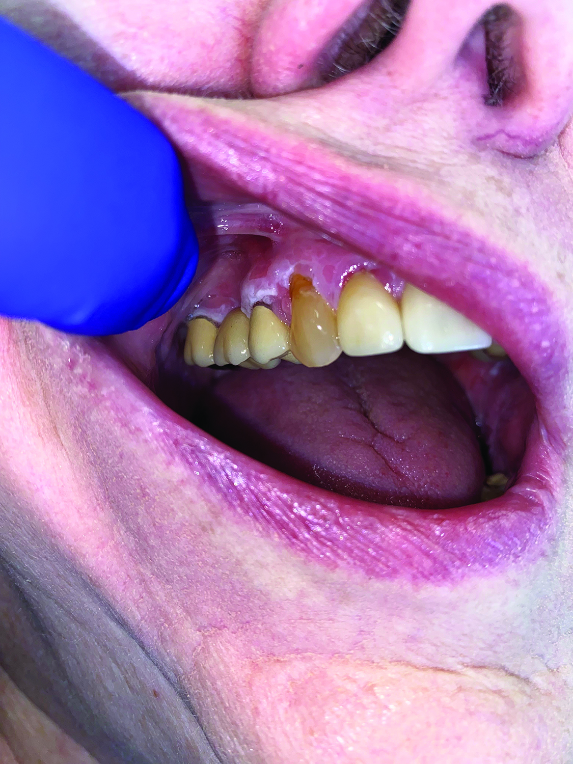
Fig. 2
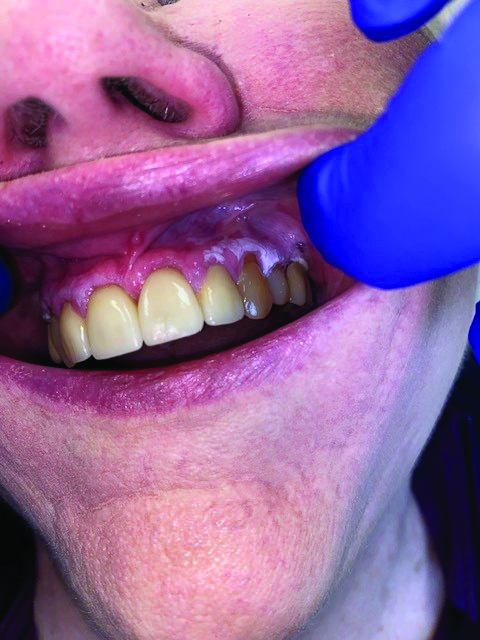
Fig. 3
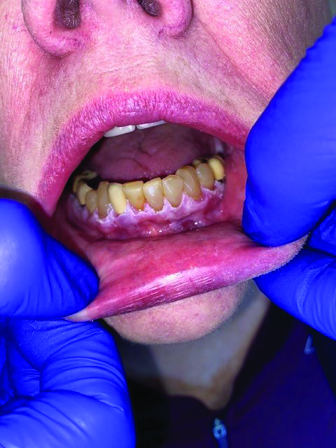
Fig. 4
The most correct diagnosis is:
A. Oral lichen planus
B. Chronic hyperplastic candidiasis
C. Proliferative verrucous leukoplakia
D. Frictional hyperkeratosis
If correct, you will be entered to win a $100 CAD Amazon gift card. **Contest end date: September 15th, 2025 at 11:59pm