Giuseppe Corinaldesi, MD, DDS, Giuseppe Lizio, DDS, et al
ackground: Mandibular second molar
(M2) periodontal defects following third molar (M3) removal in
high-risk patients is a clinical dilemma for clinicians. This study
compared the healing of periodontal intrabony defects at the distal
surface of the mandibular M2 using resorbable and non-resorbable
membranes.
Methods: Eleven patients with bilateral pocket
depth = 6 mm distal to mandibular M2 and intrabony defect = 3 mm,
related to total impaction of M3, were treated with third molar
extraction, covering the surgical bone defect with a resorbable collagen
barrier on one side and a non-resorbable polytetrafluoroethylene
(e-PTFE) barrier contralaterally. The probing pocket depth (PPD),
probing attachment level (PAL), M2 molar mobility, and grade of
furcation probing were evaluated preoperatively,and 3,6, and 9 months
postoperatively. Intraoral periapical radiographs were taken
preoperatively, immediately and at 3 and 9 months postoperatively.
Results:
Both treatment modalities were successful. At 9 months, the mean PPD
reduction was 5.2±3.9 mm for resorbable sites and 5.5±3.0 mm for
non-resorbable sites; the PAL gain was 5.9±3.3 and 5.5±3.4 mm,
respectively. The outcome difference between the two sites for both PPD
and PAL did not differ statistically (p > 0.05) at any assessment time (3, 6, 9 months).

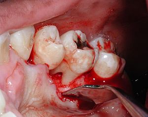
Image via Wikipedia
Conclusions:
Resorbable collagen membranes in guided tissue regeneration treatment
of intrabony defects distally to mandibular M2 obtained the same marked
PPD reduction and PAL gain as non-resorbable e-PTFE membranes following
third molar extraction.


