Anterior aesthetic prosthetics play a vital role in restoring a patient’s appearance and confidence. Achieving a seamless blend between prosthetic restorations and natural dentition requires precise shade matching, a process that remains a significant challenge in clinical dentistry. Various visual and instrumental methods have been employed to assist clinicians in matching the color of synthetic materials to natural teeth.
Despite advancements in CAD-CAM technology and digital dentistry, shade matching continues to be a complex, multi-step process. Clinicians must not only determine the correct shade but also communicate this information effectively to the dental laboratory, where technicians—who often do not see the patient—must replicate the natural dentition using available data.
This article outlines a streamlined workflow incorporating gray card calibration, DSLR photography, and Miyo stain application to enhance shade communication and improve aesthetic outcomes.
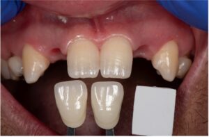
To ensure accuracy in shade selection, we prioritize shade determination at the beginning of the appointment to minimize dehydration-induced color variations. A standardized approach using a shade guide helps establish an initial reference point (Fig. 1). Additionally, capturing images with multiple adjacent shade tabs enhances both clinical documentation and laboratory interpretation (Fig. 2).
Fig. 1
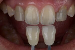
Fig. 2
Gray card calibration for standardized shade matching:
While photographs with two or more shade tabs provide a useful visual reference, this method remains subjective and dependent on the ceramist’s experience and intuition. The absence of quantifiable values introduces variability in shade reproduction.
To address this, incorporating a gray card enables digital shade verification, offering a calibrated reference point for consistent color matching. This method allows the ceramist to confirm shade accuracy before delivering the final restoration (Fig. 3).
Fig. 3
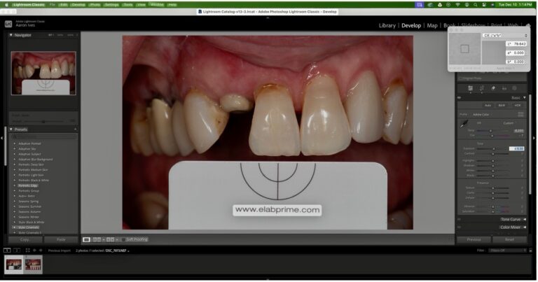
The gray card is an essential tool in digital dental photography, used to correct color distortion caused by variable lighting conditions. When taking intraoral photographs, the gray card should be placed adjacent to the teeth, and post-processing software (Lightroom or Photoshop) should be used to adjust the white balance to the card’s standardized CIE Lab* values:
• L* = 79
• a* = 0
• b* = 0
By implementing this calibration, we ensure color accuracy across different lighting conditions and provide the ceramist with a consistent reference (Fig. 4).
Fig. 4
Case study 1
Post-orthodontic replacement of congenitally missing lateral incisors
An 18-year-old male was referred for the replacement of congenitally missing upper lateral incisors following orthodontic treatment. Single-wing Maryland bridges were chosen as the treatment modality. To ensure optimal soft tissue development, the orthodontic retainer was used to compress the pontic sites.
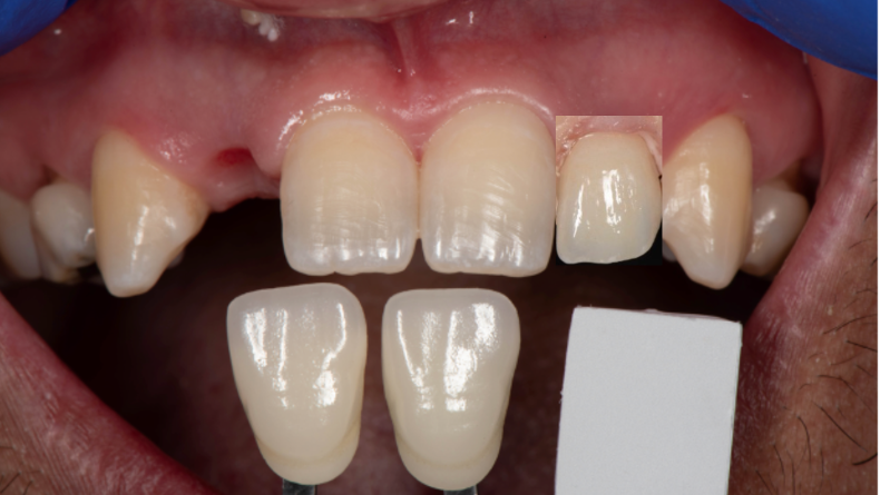
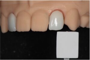
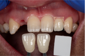
For shade documentation, a customized reference was created by attaching a square segment of the gray card to a shade tab holder. This allowed for direct incorporation of a standardized calibration point within the clinical photograph (Fig. 5).
Fig. 5
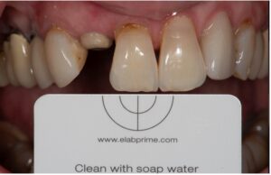
The clinical photograph was uploaded into Lightroom for color calibration, ensuring accurate L*a*b* value adjustments based on the gray card reference. The restoration was digitally designed using Exocad, milled from zirconia, and sintered. A laboratory photograph was taken using the same gray card segment to maintain color calibration consistency (Fig. 6). The photograph is cropped to capture the restoration only, the image is superimposed using power point or keynote over the clinical photo that have been previously calibrated to the gray card to compare the shade.
Fig. 6
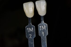
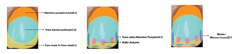
A BL2 zirconia block was used to fabricate the restoration. The initial digital try-in revealed a noticeable color discrepancy (Fig. 7), which was expected, given that the closest-matching shade tabs were 1M1 and 1M2. To refine the final shade, a shade mapping process was implemented to guide the application of Miyo stains (Fig. 8).
Fig. 7
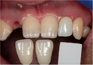
Fig. 8
Miyo stains are highly effective in fine-tuning color and translucency effects in anterior restorations. Their high translucency and excellent color saturation allow for detailed shade customization, enabling a natural replication of enamel and dentin characteristics. By layering the stains over bisque-fired prosthetics and re-firing them multiple times, we achieve precise shading, depth, and translucency that mimic natural teeth. A digital shade verification is done prior to intraoral try in of the restoration (Fig. 9) following the same steps previously described.
Fig. 9
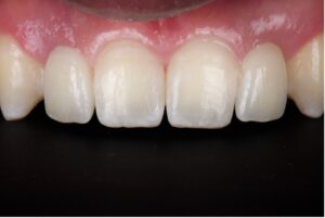
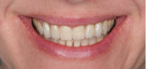
Once the digital try in confirms a satisfactory shade match, the restoration can be tried in intraorally for final confirmation and approval of the patient for final bonding (Fig. 10). These restorations are single wing Maryland bridges and are bonded following the APC protocol to bond zirconia.
Fig. 10
Case study 2
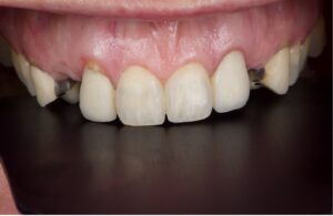
A female patient with a history of congenitally missing laterals and canine substitution done over 20 years ago presented to our office for replacement of the existing restorations for a more aesthetic solution. After a second round of orthodontic treatment the patient presents with missing teeth on the canine position (canines remained in the lateral incisor sites) and old unesthetic pbm restorations on the abutment teeth. The maxillary central incisors on this patient present with significant variations in chroma, texture and incisal effects (Fig. 11).
Fig. 11
After undergoing orthodontic treatment two times and having bridges done in the past the patient understands the value of preserving the natural dentition and does not want to have any work done on the central incisors.
Monolithic zirconia bridges from 1.2 to 1.4 and 2.2 to 2.4- were fabricated following the described protocol in this article for shade matching and Miyo stain application. The result is high strength (monolithic) restorations that blend in seamlessly with the intact natural central incisors (Fig. 12).
Fig. 12
Conclusion
By integrating gray card calibration, DSLR photography, and Miyo stains, we have established a systematic workflow that enhances shade communication and prosthetic aesthetics. This approach minimizes subjectivity, ensuring predictable and reproducible results in anterior restorations. The combination of digital shade mapping, laboratory calibration, and ceramic staining techniques allows clinicians and ceramists to achieve highly accurate and lifelike restorations consistently (Figs. 11 and 12).
By refining traditional shade-matching protocols with quantifiable digital standards, we can enhance the reliability of color reproduction and improve overall prosthetic outcomes. 
Oral Health welcomes this original article.
References
- Rondón LF, Ramírez R, Pecho OE. Comparison of visual shade matching and photographic shade analysis. J Esthet Restor Dent. 2022 Mar;34(2):374-382.
- Saygılı S, Albayrak B, Sülün T. Effect of different instrumental techniques and clinical experience on shade matching. J Prosthodont. 2024 Jun 13.
- Hein S, Zangl M. The use of a standardized gray reference card in dental photography to correct the effects of five commonly used diffusers on the color of 40 extracted human teeth. Int J Esthet Dent. 2016 Summer;11(2):246-59. PMID: 27092350.
- Blatz MB, Alvarez M, Sawyer K, Brindis M. How to Bond Zirconia: The APC Concept. Compend Contin Educ Dent. 2016 Oct;37(9):611-617
About the authors

Sangkyun “Kayden” Oh, CDT, graduated from a dental technology college in 2003 in South Korea. Starting in 2012, he managed ‘OK Line Dental Lab,’ and in 2018, immigrated to Canada. Kayden currently is partner and ceramist of CEYX Dental Lab in Winnipeg, Manitoba.

Dr. Viquez is a prosthodontist working in private practice in Winnipeg, Manitoba. He graduated from dental school in Costa Rica and did his prosthodontic residency program in Louisiana State University, where he also completed a one-year surgical dental implant fellowship. Dr Viquez is a member of multiple local study clubs and dedicates some of his time to teach at the Dr. Gerald Niznick College of Dentistry.