It has long been a goal of dental clinical and material science to find a less technique sensitive approach to the posterior direct resin restorative process. Unlike amalgam, the technique “insensitive” patrician of direct restoratives, successful placement of composite resins requires many more steps and exacting technique to achieve clinically acceptable results. So…what does an “ideal” direct resin restorative material look like? When dentists (end users) are asked about what improvements they would like to see in posterior direct resin materials, these are some of the responses they have given:
1) Ability for bulk filling (increased depth of cure)
2) Ease of delivery or placement
3) Better adaptation to the cavity preparation
4) Consistent interproximal contacts
5) Long-term predictability or reliability
6) Elimination of voids in the restoration
7) Strength to withstand functional (occlusal) forces
8) Minimal to no shrinkage upon polymerization
Since the introduction of composite resins in the mid 1980’s, composite manufacturers have made large strides in accomplishing many of these “wishes”. However, there is still a way to go before we arrive at the “ideal” direct resin restorative…or is there?
Incremental versus bulk fill technique for composite placement: A clinical dilemma
Traditionally, clinical placement of composite resins has been done using an incremental placement technique. Because of polymerization shrinkage, as well as the inability to light cure composite materials beyond a certain depth, it has been generally recommended to place composite resin in increments of two millimeters or less. The question is with advances in polymer chemistry, photo activation, and curing light technologies that we currently see with today’s composite resin materials, is this still true? Several studies have been done, some as early as 2001, that compare incremental versus bulk fill placement of composite that show that there is no difference in cuspal deflection or marginal integrity when comparing techniques of placement. The main clinical issue with bulk fill placement is depth of cure, or the inability of the curing light in a deep preparation to reach the photo initiator in the composite material to cause polymerization of the material. It is also important to note that directional curing from the buccal and lingual (palatal) aspects of a Class II preparation after removal of the matrix help increase the ability to cure composite at the gingival margin of the proximal box in a Class II restoration.1-3
Incremental placement, regardless of the classic “2mm incremental approach” of bulk placement of a “dentin material” followed by a separate “capping” layer of conventional composite has been shown that microleakage and inaccurate adaptation of the restorative material can lead to the ingress of recurrent decay and ultimate restoration failure.4-6
Bulk fill flowable materials as “dentin replacements” for posterior composite restorations
In 2010, bulk fill flowable composites were introduced to the dental marketplace, the first being Surefil SDR (Smart Dentin Replacement) by Dentsply Caulk. Since then, many other bulk fill flowable composites have followed. These materials are indicated for use as a bulk fill base (dentin replacement) beneath posterior composite restorations and can be placed in a single increment up to 4mm in depth. Being able to place that amount of material in a single increment is a significant time saver, and while the concept sounds quite simple, there are several important requirements a material must meet for this particular indication. According to the manufacturers, these include 1) increased depth of cure, 2) a viscosity that will readily adapt to the internal walls of the cavity without the need for manipulation of the material, and 3) low polymerization shrinkage stress. Because of their transparent nature, and decreased percentage of filler particles, most bulk fill flowable composites will require a conventional nano hybrid composite material to be placed as the “enamel-capping layer.”7
A bioactive component to bulk fill restorative materials
What exactly does “bioactivity” mean in dentistry today? In addition to the effect of fluoride on healthy hydroxyapatite, making enamel less susceptible to demineralization due to the acid byproducts of bacterial metabolism, the ability of a material to contribute calcium and phosphate ions to help rebuild demineralized dentin is the new paradigm. These new “smart” bioactive dental materials react to pH changes in the oral environment to elicit changes in the material and the tooth as well. Hence, “biomineralization” refers to the exchange of calcium and phosphate ions within the tooth substance forming new hydroxyapatite, and in many cases, repairing existing demineralized hydroxyapatite. Bioactivity also includes the precipitation of hydroxyapatite crystals on the surface of the material in the presence of moisture (saliva). This phenomenon has the potential to keep restoration margins sealed for extended periods of time due this mineral-like deposition at the tooth-restorative interface.10-12
The advantages of bioactive restorative materials are clear after reading the above discussion regarding bioactivity and the effects of rebuilding damaged tooth structure. Having restorative materials that can protect, as well as repair and help seal the marginal gap that exists around all dental restorations, can help preserve the natural tooth and extend the life of the dental restoration. Activa Restorative (Pulpdent Corporation) is one such restorative material. It is particularly useful for patients with high caries indices where getting control of the problem can be difficult. Acidic oral environments can create extensive damage to teeth, particularly if a patient doesn’t know which end of the toothbrush has the bristles! Pediatric patients and primary teeth have their own sets of issues as well. Finishing a procedure quickly in a less than ‘moisture-free” environment makes using traditional composites and adhesives very difficult on pediatric patients.
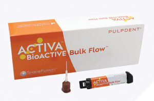
With the recent introduction of Activa Bulk Fill (Pulpdent Corporation) brings us closer to that “ideal restorative material” that has been envisioned for years. In addition to satisfying all the improvements that dentists have asked for in a posterior restorative mentioned at the beginning of this article, Activa Bulk Fill has all the bioactive benefits of Activa Bioactive Restorative. And like Activa Bioactive Restorative, it is a dual cure material which eliminates any depth of cure issues that may exist in larger cavities. Dual cure capability offers an advantage in deeper cavities where it can be difficult for curing lights to reach and completely cure the bottom of the restoration. The addition of bioactive properties to a bulk-fill restorative material gives long-term benefits to the patient helping to defend against secondary caries by decreasing biofilm accumulation, remineralization of demineralized tooth structure, and maintenance of restorative marginal seal by facilitating the deposition of hydroxyapatite crystals on the surface of the restorative material.13 Another advantage over the original Activa Bioactive Restorative material is the new syringe delivery, which greatly simplifies delivery and placement. In this case study, we will look at how Activa Bulk Fill (Pulpdent Corporation) utilizes self-leveling properties, has minimal shrinkage stress due to Modulus (a patented rubberized resin technology), and provides excellent single-shade aesthetics making it a superior choice for the direct restoration of posterior teeth. It is for these reasons Activa Bulk Fill (Fig.1) was chosen for this case.
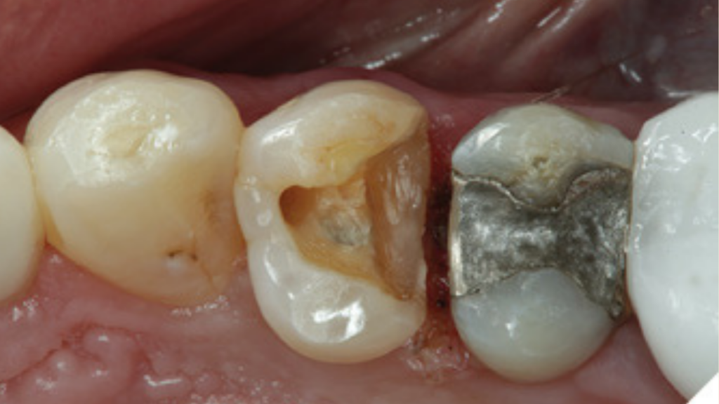
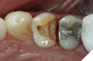
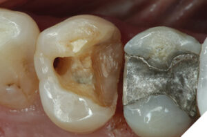
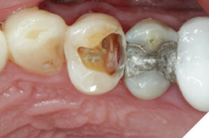
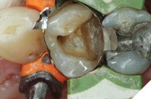
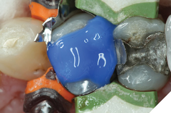
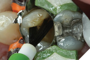
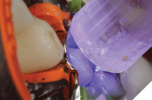
The patient, a 27-year-old male, presented with a fractured composite restoration with recurrent decay diagnosed by radiograph on the distal surface of tooth # 24. Although a full coverage indirect restoration was recommended for this tooth, the patient opted for a direct restoration at this time. A preoperative occlusal view of tooth # 24 is shown in Figure 2. Figure 3 shows that after initial removal of the old restorative material except for the marginal ridge portion, it is evident that the gingival wall of the original cavity preparation was never properly sealed. The remainder of the old restorative material is removed, and a diode laser is used to further expose the gingival extent of the preparation. This will help ensure proper seal of the gingival margin when the matrix is placed since the tissue will not impede its placement (Fig. 4). Caries was also observed on the mesial aspect of the preparation running under the mesial marginal ridge toward the proximal surface below the contact area (Fig. 5). A sectional matrix and ring have been utilized on both the mesial and distal surfaces of the Class II preparation to contain the restorative material (Fig. 6). Precise placement of the matrix band at the cavosurface margins and marginal ridge to confine the restorative material is very important to prevent marginal overflow and excessive contouring after placement. Next, implementing “total etch” technique, the preparation is etched for 15 seconds (Fig. 7), rinsed, then dried being careful not to over desiccate the dentinal surface. A universal adhesive is placed with a brush (Fig. 8), air thinned to evaporate the solvent, then light cured per manufacturer’s instructions (Fig. 9).
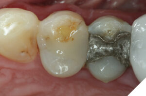
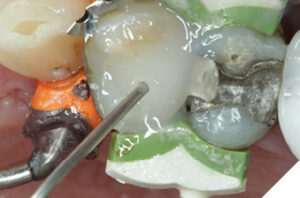
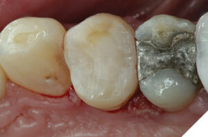
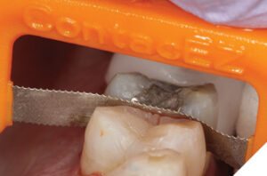
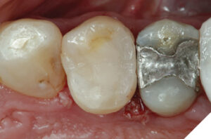
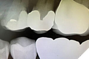
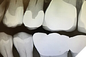
Activa Bulk-fill is then delivered to the preparation in one bulk increment (Fig. 10). The viscosity is such that the material readily flows and accurately fills the entire preparation with intimate adaptation to the cavity walls. The ability of the material to “self-level” helps limit the potential of overfilling the cavity preparation during placement. Next, the material is light cured based on manufacturer’s instructions. Figure 11 shows placement of Activa Bulk-fill after it is placed, and light cured. Notice the cavity was not overfilled and there is very little excess due to the precise fit of the sectional matrix band. This will simplify and speed up the finishing process of the restoration. Figure 12 shows use of an abrasive device (ContacEZ: Directa USA) to refine proximal contour and contact area. A needle-shaped diamond composite finishing bur (mosquito bur) is used to accentuate anatomic form and make any minor occlusal adjustments. Rubber abrasives are then used to obtain a final polish to the restoration (A.S.A.P. – All Surface Access Polishers: Clinical Research Dental). After placement and curing of a composite surface sealant (Seal-N-Shine: Pulpdent Corporation), the final Activa Bulk-fill restoration on tooth number 24 is shown in Figure 13. Notice how the single-shade aesthetics makes these restorations disappear into the natural tooth structure. Figure 14 is a preoperative bite wing x-ray of tooth number 24 showing the extent of the recurrent decay on the distal aspect apical to the old composite restoration. Figure 15 is a postoperative bite wing x-ray showing the completed placement of the Activa Bulk-fill restoration on tooth number 24. It is important to note that the mesial aspect of the amalgam restoration on tooth number 25 was recontoured prior to placement of the Activa Bulk-fill restoration on tooth number 24 to eliminate the overhang and create a more ideal interproximal embrasure. This procedure should always be done, as needed, on adjacent restorations (teeth) prior to new restorative placement to create more physiologic tooth contours in the gingival embrasure spaces.
Epilogue
We are now closer than ever before to an “ideal” direct restorative material. Precise placement is now more predictable using a single increment, dual cure restorative that also provides long term bioactive benefits. Extending the life of direct restorative materials before requiring replacement has the potential benefit of helping patients keep their natural teeth longer without the need for more extensive restoration or replacement by implants.
 References
Sauro S, Pashley, DH, Strategies to stabilise dentine-bonded interfaces through remineralising operative approaches – state of the art, Int J Adhesion and Adhesives 2016.
Campodonico CE, Tantbirojan, D, Olin PS, Versluis A, Cuspal deflection and depth of cure in resin based composite restorations filled by using bulk, incremental, and trans tooth illumination techniques, JADA, 142 (10), October, 2011, pp. 1176-1182.
Rees JS, et. al., A reappraisal of the incremental packing technique for light cured composite resins, J of Oral Rehab 2004; 31: 81-84.
Flury S, Hayoz S, Peutzfeldt A, Hüsler J, Lussi A. Depth of cure of resin composites: is the ISO 4049 method suitable for bulk fill materials? Dent Mater. 2012 May; 28(5):521-8.
Ilie N, Bucuta S, Draenert M. Bulk-fill resin-based composites: an in vitro assessment of their mechanical performance. Oper Dent. 2013 Nov-Dec; 38(6):618-25.
Juloski J, Carrabba M, Aragoneses JM, Forner L, Vichi A, Ferrari M. Microleakage of Class II restorations and microtensile bond strength to dentin of low-shrinkage composites, Am J Dent. 2013 Oct;26(5):271-7.
Van Ende A, De Munck J, Van Landuyt KL, Poitevin A, Peumans M, Van Meerbeek B. Bulk-filling of high C-factor posterior cavities: effect on adhesion to cavity-bottom dentin. Dent Mater. 2013 Mar; 29(3):269-277.
Roggendorf MJ, Krämer N, Appelt A, Naumann M, Frankenberger R. Marginal quality of flowable 4-mm base vs. conventionally layered resin composite. J Dent. 2011 Oct;39(10):643-7.
Yapp R, Powers JM. Depth of Cure of Several Composite Restorative Materials. Dental Advisor Res Rpt 33:1. February 2011.
Alrahlah, A.; Silikas, N.; Watts, D.C. Post-cure depth of cure of bulk fill dental resin-composites. Dental Materials. Feb 2014, Vol. 30 Issue 2, p149-154.
Hench LL, et al.J. Direct chemical bond of bioactive glass ceramic materials to bone and muscle, Biomed. Mater. Res., 2 (1972) vol 7, # 3, pp. 25-42.
McCabe JF, et al, Smart materials in dentistry, Aust Dent 2011 June; 56 Suppl 1pp.3-10.
Jefferies SR. Preliminary evidence that bioactive cements occlude artificial margin gaps, J Esthet Restora Dent 2014;26(1): 14-26.
About the author:

Dr. Robert A. Lowe graduated Loyola University magna cum laude. He is Assistant Professor in the Department of Oral Rehabilitation, Medical University of South Carolina and a clinical evaluator of materials and products. He lectures internationally and publishes extensively on esthetic and restorative dentistry. Dr. Lowe has been awarded Fellowships in the AGD, ICD, ADI, ACD, ASDA, IADFE and DiplABAD, and received the 2004 Gordon Christensen Outstanding Lecturers Award. A founding member of the World Clinical Laser Institute, Dr. Lowe maintains a part-time private practice in Charlotte, North Carolina.