
Missing maxillary lateral incisors are one of the most common congenitally missing tooth conditions that exists and occurs in approximately 1-3% of the population.1 It is the most common missing tooth after the second premolar when excluding third molar agenesis and has been reported to be more prevalent in females and occurs more bilaterally than in unilaterally.1-4 Largely, the cause of dental agenesis is due to genetics, however environmental factors such as trauma or radiation may lead to dental agenesis as well.5
Patients who present with congenitally missing laterals face a functional and aesthetic problem at a young age. This can affect their self-esteem as well as their social relationships, especially during adolescence.6 Multiple treatment options are available for patients whom have congenitally missing lateral incisors – these options include canine substitution, traditional fixed partial dentures, resin bonded fixed partial dentures, as well as single implant crowns.7 Often, orthodontics treatment for space redistribution is also necessary before prosthetic treatment can be completed. Choosing the right treatment option is based on multiple factors including patient age, psychology, functional risk, and aesthetic demands. The single implant crown could be considered by many the most ideal treatment option – it involves no preparation of adjacent teeth, ease of oral hygiene, as well as the ability to achieve high aesthetic and functional demands and is widely supported in the literature8.
For patients with congenitally missing lateral incisors, an important aspect to consider prior to implant placement is patient age. Often these patient’s want some form of prosthetic treatment around adolescence due to self-image and social factors9. However, placement of implants at an early age may cause complications in the future and has to be weighed during the treatment planning process.10,11 Implant placement should ideally only be performed after completion of facial growth as dental implants are fused to the bone and any potential growth will not move the implant itself. An implant placed when growth has not completed will be in infraocclusion and can become a very difficult aesthetic problem.10 Currently there is no consensus of age to place implants in adolescents – studies have shown growth continues for females until 17 and men until 219,12,13 however there are reported instances of growth continuing well beyond those years.14 Management of cases where implants were placed at an early age and continuous maxillary growth has occurred can be very challenging. Prevention can be attempted with a fixed retainer, however if growth were to continue, there is potential for an open occlusal relationship as the implants prevent the adjacent teeth from growing.15 Modifying the implant restoration and neighbouring teeth is often chosen as a treatment option as it is the least invasive.15 In severe cases, surgical repositioning of the implant has been documented through distraction osteogenesis.16
Case Report
Patient info
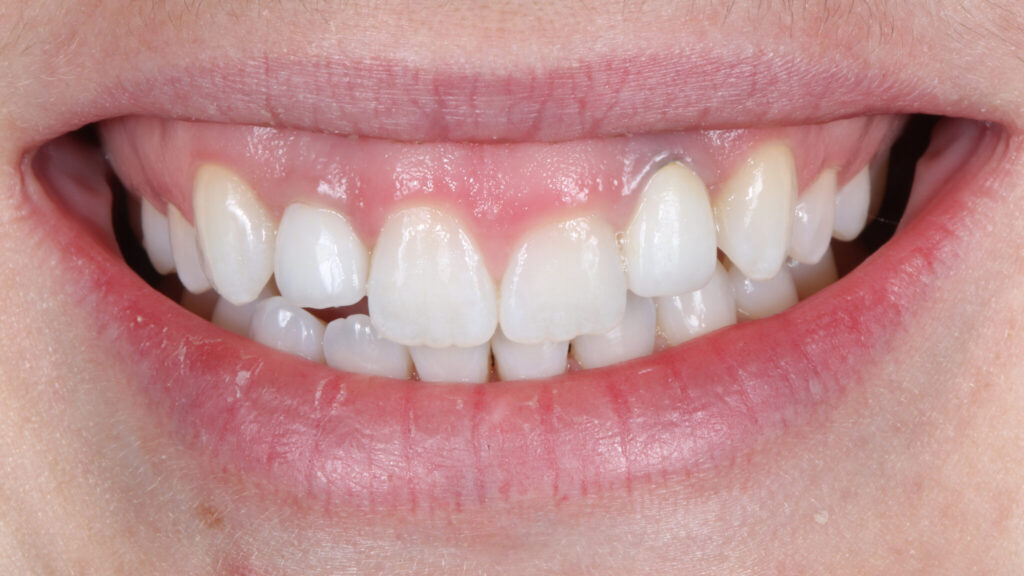
A 27-year-old female presented to our office with a chief concern of disliking the appearance of her anterior teeth, especially the gum levels. The patient had congenitally missing lateral incisors and had previously had orthodontics to align her teeth and create space for two implants in the lateral sites. The two implants (Nobel Active 3.0, Nobel BioCare) were placed approximately 10 years ago and were subsequently restored with porcelain-fused-to-metal cemented crowns.
Upon examination, there is some greyness to the original implant crowns due to the show-through of the metal underneath as well as the colour being a bit higher in value than the rest of her dentition. The gingival levels of her teeth and implants were uneven – notably the gingiva of implant #12 is much more apical compared to tooth #11. Luckily for this patient, there was passive altered eruption on her maxillary central incisors – which indicates that the gingival level can be pushed apically and the remaining tooth can be exposed with crown lengthening treatment. A combination of new implant crowns and crown lengthening was treatment planned for this patient. It was noted that there was a possibility that veneers were required in order to achieve a harmonious result after crown lengthening. If crown lengthening had resulted in root exposure, or the proportions of the teeth were suboptimal, then veneers would be recommended in order to achieve a satisfying aesthetic result.
Crown lengthening was completed by a periodontist, followed by 12 weeks of healing and stabilization of the gingival levels. The patient then returned to have new implant crowns fabricated. In order to facilitate removal of the old cemented crowns, holes were drilled through the crowns in order to access the screws. Upon removal of her original implant crowns, the implant positions were evaluated. The #12 implant positioning goes straight through the incisal edge; however, the #22 implant is severely buccally proclined.
Two new screw-retained temporary restorations were fabricated in an attempt to achieve better emergence contours and achieve some downward gingival growth. Note the greyness of the temporary as the buccal surface was too thin to block-out the colour of the titanium temporary cylinder.
The final implant crowns were designed with custom milled gold-anodized titanium abutments and cemented zirconia crowns. Due to the type and size of the implant which was placed (Nobel Active 3.0; Nobel BioCare), an angled solution was not available. This also emphasizes that all prosthetic possibilities should be considered before implant surgery in order to know your prosthetic options afterwards. In situations where the angulation of the implant may not be perfect and an angled screw may be required, an implant system should be selected in order to accommodate this.
The soft tissue contours were impressed and given to the lab to copy the emergence profile developed by the temporaries. Final impressions were made and custom abutments fabricated. The gold anodized abutment benefits the colour of both the tissues and the crown as the amount of restorative space buccally was limited. After the final abutments were torqued in, the implant crowns were cemented using a permanent cement.
Discussion
Treating patients with implants in the aesthetic region placed at an early age present many aesthetic challenges. Often, cases like these involve compromise due to the location of the implants as they cannot be moved. The goal of restorative treatment is to try to camouflage and blend the implant crowns to the rest of the smile as much as possible. Not every case is as lucky enough to have altered passive eruption and require crown lengthening surgery on the remaining dentition to be able to even the gingival levels. In cases where teeth are fully erupted, crown lengthening surgery can cause root exposure and veneers may be required for both root coverage and overall aesthetics.
Another factor that should be emphasized is the importance of knowing the prosthetic options for the implants which are being placed. Had the implant system had an angled solution available, then at least the #12 implant could have been screw retained for ease of retrieval in the future and no cementation process been required. Interdisciplinary care is also imperative in treating cases similar to this. Having input from a periodontist, oral surgeon, as well as an orthodontist can help treatment plan these cases and achieve the most optimal results possible.
Fig. 1

Fig. 2
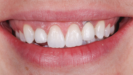
Fig. 3

Fig. 4
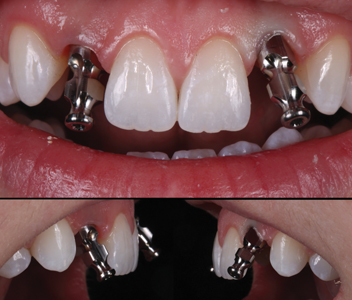
Fig. 5

Fig. 6
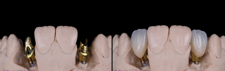
Fig. 7

Conclusion
The use of dental implants to treat congenitally missing laterals is a great treatment option, however care needs to be taken on case selection and implant timing. In cases where implants are placed too early, situations involving implant infraocclusion may arise and will have a large impact on the aesthetics of a patient’s smile. Treatment of these cases can be complex and vary greatly. For this patient, we were able to achieve a satisfactory result through the combination of periodontal crown lengthening surgery and new prosthetic implant crowns.
Oral Health welcomes this original article.
References
- Kavadia, S., Papadiochou, S., Papadiochos, I., & Zafiriadis, L. (2011). Agenesis of maxillary lateral incisors: a global overview of the clinical problem. Orthodontics : the art and practice of dentofacial enhancement, 12(4), 296–317.
- Fujita, Y., Science, S. F., Hidaka, A., Science, S. F., Nishida, I., Science, F., … Science, S. F. (2009). Developmental Anomalies of Permanent Lateral Incisors in Young Patients. J Clin Pediatr Dent, 33(3), 211–216.
- Altug-Atac, A. T., & Erdem, D. (2007). Prevalence and distribution of dental anomalies in orthodontic patients. American Journal of Orthodontics and Dentofacial Orthopedics, 131(4), 510–514. https://doi.org/10.1016/j.ajodo.2005.06.027
- Polder, B. J., Van’t Hof, M. A., Van Der Linden, F. P. G. M., & Kuijpers-Jagtman, A. M. (2004). A meta-analysis of the prevalence of dental agenesis of permanent teeth. Community Dentistry and Oral Epidemiology, 32(3), 217–226. https://doi.org/10.1111/j.1600-0528.2004.00158.x
- Papageorgiou, S. N., Eliades, T., & Hämmerle, C. H. F. (2018). Frequency of infraposition and missing contact points in implant-supported restorations within natural dentitions over time: A systematic review with meta-analysis. Clinical Oral Implants Research, 29(May), 309–325. https://doi.org/10.1111/clr.13291
- Rakhshan, V., & Meline. (2015). Concerning the Etiology , Prevalence, Risk Factors , Patterns and Treatment. Dental Research Journal, 12(1), 25–27.
- Krassnig, M., & Fickl, S. (2011). Congenitally missing lateral incisors-A comparison between restorative, implant, and orthodontic approaches. Dental Clinics of North America, 55(2), 283–299. https://doi.org/10.1016/j.cden.2011.01.004
- Branzén, M., Eliasson, A., Arnrup, K., & Bazargani, F. (2015). Implant-Supported Single Crowns Replacing Congenitally Missing Maxillary Lateral Incisors: A 5-Year Follow-Up. Clinical Implant Dentistry and Related Research, 17(6), 1134–1140. https://doi.org/10.1111/cid.12233
- Mankani, N., Chowdhary, R., Patil, B. A., Nagaraj, E., & Madalli, P. (2014). Osseointegrated dental implants in growing children: A literature review. Journal of Oral Implantology, 40(5), 627–631. https://doi.org/10.1563/AAID-JOI-D-11-00186
- Klinge, A., Tranaeus, S., Becktor, J., Winitsky, N., & Naimi-Akbar, A. (2021). The risk for infraposition of dental implants and ankylosed teeth in the anterior maxilla related to craniofacial growth, a systematic review. Acta Odontologica Scandinavica, 79(1), 59–68. https://doi.org/10.1080/00016357.2020.1807046
- Op Heij, D. G., Opdebeeck, H., Van Steenberghe, D., & Quirynen, M. (2003). Age as compromising factor for implant insertion. Periodontology 2000, 33, 172–184. https://doi.org/10.1046/j.0906-6713.2003.03314.x
- Bohner, L., Hanisch, M., Kleinheinz, J., & Jung, S. (2019). Dental implants in growing patients: a systematic review. British Journal of Oral and Maxillofacial Surgery, 57(5), 397–406. https://doi.org/10.1016/j.bjoms.2019.04.011
- Holmes, J. D. (2013). Considerations in Dental Implant Placement in the Young Patient: A Surgeon’s Perspective. Seminars in Orthodontics, 19(1), 24–36. https://doi.org/10.1053/j.sodo.2012.10.001
- Bernard, J. P., Schatz, J. P., Christou, P., Belser, U., & Kiliaridis, S. (2004). Long-term vertical changes of the anterior maxillary teeth adjacent to single implants in young and mature adults: A retrospective study. Journal of Clinical Periodontology, 31(11), 1024–1028. https://doi.org/10.1111/j.1600-051X.2004.00574.x
- Zitzmann, N. U., Arnold, D., Ball, J., Brusco, D., Triaca, A., & Verna, C. (2015). Treatment strategies for infraoccluded dental implants. Journal of Prosthetic Dentistry, 113(3), 169–174. https://doi.org/10.1016/j.prosdent.2014.08.012
- Bousquet, P., Barthélemi, S., Artz, C., & Delsol, L. (2021). The application of orthodontic bone stretching for correcting malpositioned dental implants. Head and Face Medicine, 17(1), 1–11. https://doi.org/10.1186/s13005-021-00294-y
About the Author

Dr. Max Li is a Board Certified Specialist in Prosthodontics and a Fellow of the Royal College. Dr. Li focuses his practice around creating a healthy and functional dentition along with esthetic smiles. He currently maintains a full-time specialty practice in Toronto and teaches as a clinical instructor at the University of Toronto Faculty of Dentistry.


